Submit or Track your Manuscript LOG-IN

An Abattoir-Based Study on the Prevalence of Hydatidosis Infestation and Fertility of Hydatid Cysts in Slaughtered Herbivores (Food Animals) in Dhamar Province-Yemen.

An Abattoir-Based Study on the Prevalence of Hydatidosis Infestation and Fertility of Hydatid Cysts in Slaughtered Herbivores (Food Animals) in Dhamar Province-Yemen.

Mohammed Naji Ahmed Odhah1,2*, Dhary Alewy Almashhadany6, Abdullah Garallah Otaifah7, Bashiru Garba5, Najeeb Mohammed Salah2, Faez Firdaus Abdullah Jesse4*, Mohd Azam Khan G.K3 
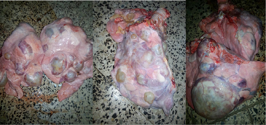
1Department of Veterinary Clinical Studies, Faculty of Veterinary Medicine, Universiti Malaysia Kelantan (UMK), Kampus Kota, Pengkalan Chepa, 16100 Kota Bharu, Kelantan, Malaysia; 2Department of Public Health, Faculty of Veterinary Medicine, Thamar Uni-versity, 87246 Dhamar, Yemen; 3Department of Veterinary Paraclinical Studies, Faculty of Veterinary Medi-cine, Universiti Malaysia Kelantan, Pengkalan Chepa, 16100 Kota Bharu, Ke-lantan, Malaysia; 4Department of Veterinary Clinical Studies, Faculty of Veterinary Medicine, Universiti Putra Malaysia 43400 UPM Serdang, Selangor, Malaysia; 5Department of Veterinary Public Health and Preventive Medicine, Faculty of Veterinary Medicine, Usmanu Danfodiyo University, Sokoto, Nigeria; 6Department of Medical Laboratory Sciences, College of Science, Knowledge University, Erbil City, Kurdistan Region Iraq; 7Department of Anatomy, School of Medical Science, Universiti Sains Malay-sia (USM), 16150 Kubang Kerian, Kelantan, Malaysia.

*Correspondence | Mohammed Naji Ahmed Odhah, Department of Veterinary Clinical Studies, Faculty of Veterinary Medicine, Universiti Malaysia Kelantan (UMK), Kampus Kota, Pengkalan Chepa, 16100 Kota Bharu, Kelantan, Malaysia; Email: naji.ao@umk.edu.my; jesse@upm.edu.my 

Figure 1

The lung of cattle showing multiple hydatid cysts of varying sizes 
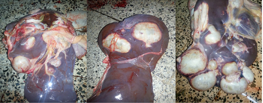
Figure 2

The liver of cattle showing multiple hydatid cysts of varying sizes 
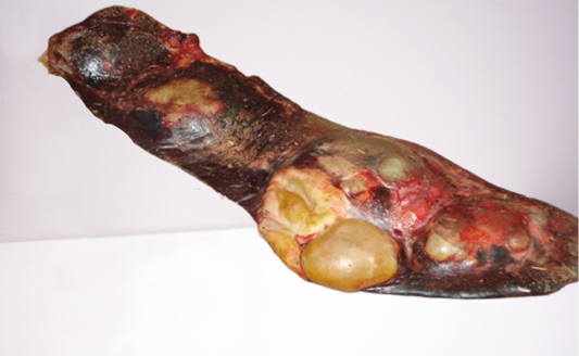
Figure 3

Spleen showing multiple hydatid cyst in the slaughtered cattle  
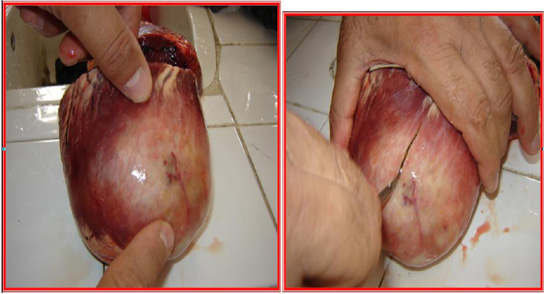
Figure 4

Showing the solitary hydatid cyst in the heart in cattle 
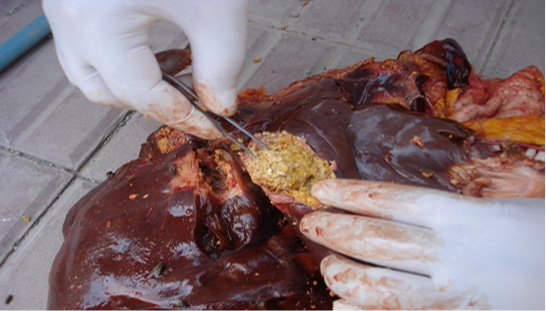
Figure 5

Shows the presence of encrusted water cysts in the liver of local cattle. 
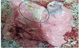
Figure 6

Indicate the presence of encrusted water cysts (encircled) in the lungs of local cattle. 
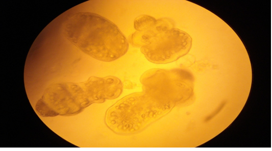
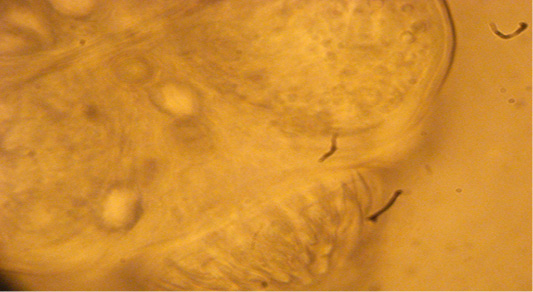
Figure 7

Showing the external shape of the spines on the tapeworm heads 

Figure 8

Shows the germinative membrane removed from a cyst taken from the liver of an infected cow. 

Journal of Animal Health and Production

October

Vol. 11, Iss. 3, Pages 234-329

Featuring

Click here for more

Subscribe Today

Receive free updates on new articles, opportunities and benefits


Subscribe Unsubscribe