Submit or Track your Manuscript LOG-IN

Bilateral Orbital Myiasis Due to Wohlfahrtia magnifica and its Treatment in a Dog

Bilateral Orbital Myiasis Due to Wohlfahrtia magnifica and its Treatment in a Dog

Amir Masoud Jafari-Nozad1*, Keyvan Samadi2, Kamran Akbarzadeh3, Hasan Bakhshi4*, Kourosh Arzamani4 and Amirsajad Jafari5
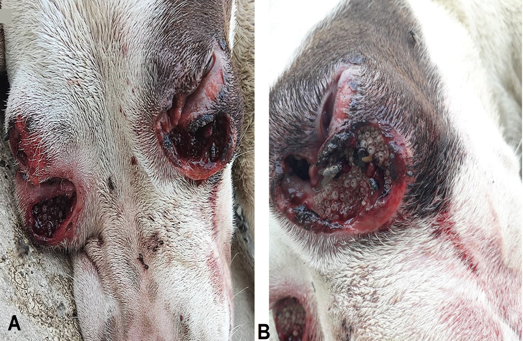
1Student Research Committee, Birjand University of Medical Sciences, Birjand, Iran.
2Oxygen Veterinary Clinic, Karaj, Iran.
3Department of Medical Entomology and Vector Control, School of Public Health, Tehran University of Medical Sciences, Tehran, Iran.
4Vector-Borne Diseases Research Center, North Khorasan University of Medical Sciences, Bojnurd, Iran.
5Department of Basic Sciences, School of Veterinary Medicine, Shiraz University, Shiraz, Iran.
 
Corresponding Author: Amir Masoud Jafari-Nozad and Hasan Bakhshi
a.sajad.jafari@gmail.com, hbakhshi89@gmail.com

Figure 1:

Clinical manifestation of orbital myiasis in an anesthetized Pitbull dog before treatment (A and B).
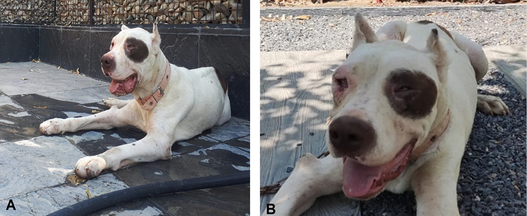
Figure 2:

Improvements in animal’s condition after three weeks of treatment.
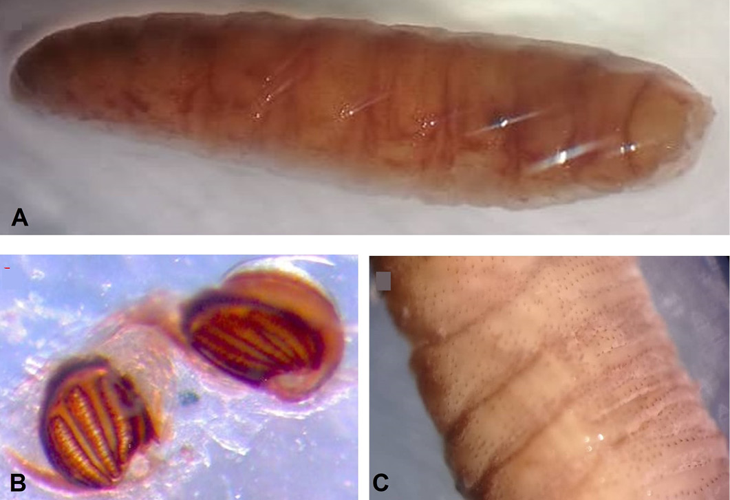
Figure 3:

Identification of larvae by morphological keys. (A) larvae, (B) spiracles in the cavity of posterior part, (C) scattered stout spines on the body surface.

Punjab University Journal of Zoology

June

Vol.38, Iss. 1, Pages 01-135

Featuring

Click here for more

Subscribe Today

Receive free updates on new articles, opportunities and benefits


Subscribe Unsubscribe