

Concentration Dependent Functions of FGF10 in Angiogenesis and Axon Guidance


Concentration Dependent Functions of FGF10 in Angiogenesis and Axon Guidance
Fang Liu*
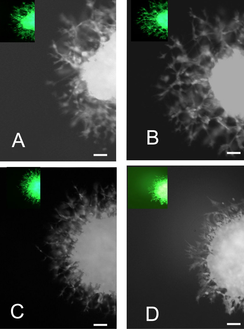
Effects of FGF10 on the outgrowth of newborn vessels. (A) Control; (B) low concentration FGF10 in culture media; (C) high concentration FGF10 in culture media; (D) low concentration FGF10 plus SU5402 in culture media. Insets in A, B, C and D Show Tie-1 expression in CAM vessels. Scale bars represent 50μm.

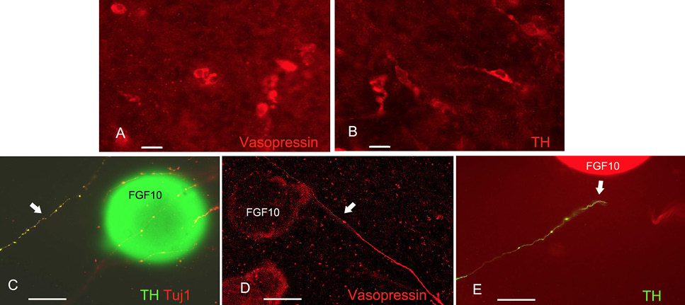
Roles of FGF10 on distinct hypothalamic axons. Vasopressin (A) and TH (B) positive neurons are present within the E4 explants. After immunolabelling with Tuj1, axons extend towards FGF10 soaked beads, but one axon (TH+) turn (arrow), rather than project into FGF10 beads (C). Vasopressin axon goes directly towards FGF10 sources (arrow, D); while TH axon turns away from FGF10 beads (arrow, E). Scale bars represent 50μm.
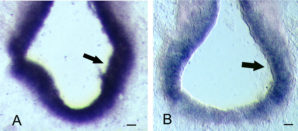
Fgfr1 (A) and Fgfr2 (B) expressions are detected throughout the hypothalamus (arrows), in both hypothalamic ventricular and mantle zone. Scale bars represent 50μm.


