


Detection of Mycoplasma bovis from Cattle Presented for Slaughter in Adamawa and Taraba States, Northeastern Nigeria
Markus I. Francis1*, Paul I. Ankeli2, Clara N. Kwanashie1, Jibril Adamu1, Lushaikyaa Allam1, Mashood A. Raji3, Godwin O. Egwu4, Flavio Sacchini5 and Massimo Scacchia5
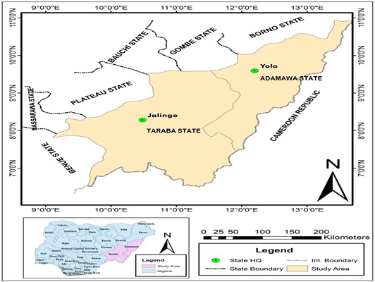
Figure 1:
Map of Adamawa and Taraba States, North-eastern Nigeria (MLS, 2010).
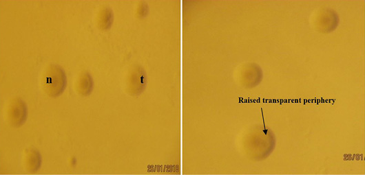
Figure 2:
Colonies of Mycoplasma bovis from lung sample on PPLO agar showing ‘fried egg’ colonies with nipples ‘n’ and raised transparent periphery ‘t’ observed under Stereomicroscope (X40).
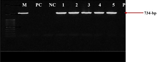
Figure 3:
PCR amplification of Mycoplasma bovis 16S rRNA gene on 1% agarose gel with primers MBOF2 and MBOR2. M: molecular marker of 50-bp; PC: positive control reference strain PG45 (734-bp); NC: negative control; Lanes 1-5: represents samples TL41, TL43, TL46, TES57 and AP106.

December 2020
Vol. 6, Iss. 2, Pages 58-149
