
Effect of Cortisone and Cortisol on Prostaglandins Production by Bovine Endometrium Around the Time of Ovulation


Effect of Cortisone and Cortisol on Prostaglandins Production by Bovine Endometrium Around the Time of Ovulation
Duong Thanh Hai1*, Tomas J. Acosta2, Tran Thi Minh Tu3
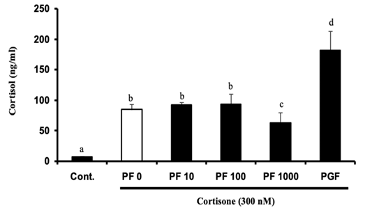
Dose-dependent effects of PF915275 (a specific inhibitor of 11β hydroxysteroid dehydrogenase type 1) on the conversion of cortisone to cortisol by bovine endometrial tissue from the follicular stage (Mean ± SEM, n=4 experiments, each performed in triplicate). Endometrial tissues were exposed to PF (0-1000 nM) or PGF (1 µM) combined with cortisone (300 nM) for 4 h. Different superscript letters indicate significant difference (p<0.05) as determined by ANOVA followed by protected least significant difference test (PLSD).
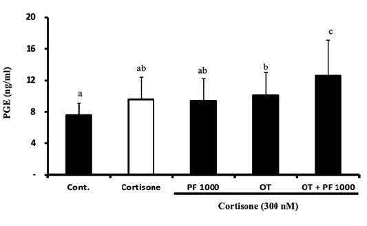
Effects of cortisone on PGE production by cultured bovine endometrial tissue at follicular stage (Mean ± SEM, n=4 experiments, each performed in triplicate). Endometrial tissues were exposed to cortisone (300 nM) alone or cortisone (300 nM) combined with oxytocin (OT, 100 nM) in the presence or absence of PF (1000 nM, PF 1000) for 4 h. Different superscript letters indicate significant difference (p<0.05) as determined by ANOVA followed by protected least significant difference test (PLSD).
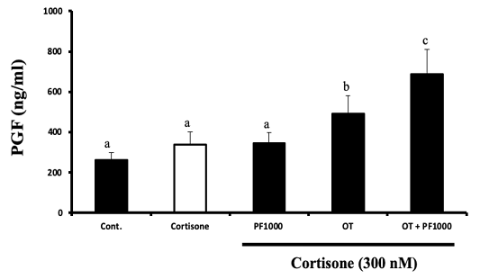
Effects of cortisone on PGF production by cultured bovine endometrial tissue at follicular stage (Mean ± SEM, n=4 experiments, each performed in triplicate). Endometrial tissues were exposed to cortisone (300 nM) or cortisone (300 nM) combined with oxytocin (OT, 100 nM) in the presence or absence of PF (1000 nM; PF 1000) for 4 h. Different superscript letters indicate significant difference (p<0.05) as determined by ANOVA followed by protected least significant difference test (PLSD).

