

Effect of Microwave Exposed Feed in Different Containers on Histological Structure of Liver and Kidney of Adult Mice


Effect of Microwave Exposed Feed in Different Containers on Histological Structure of Liver and Kidney of Adult Mice
Sajida Batool*, Sitara Shameem, Kainat Malik and Aneela Iram
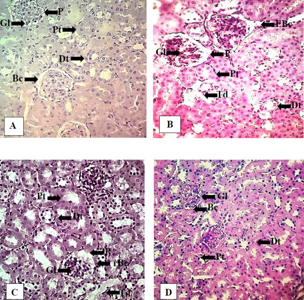
Effect of exposure of feed to microwaves in different contains on histological structure of kidney of male mice. A: Control showed well organized Bowman’s capsule (Bc) with narrow peri-glomerular spaces (P) compact glomerulus (Gl), normal proximal (Pt) and distal tubules (Dt) with slight lumen. B: Direct group showed edematous Bowman’s capsule (EBc), increased peri-glomerular space, with swollen glomeruli (Gl), tubular degeneration (Td), and dilation of proximal and distal tubules with increased lumen. C: Glass group showed ruptured Bowman’s capsule (rBc), swollen glomeruli, wide peri-glomerular space. Proximal and distal tubules showed dilation and tubular degeneration. D: Plastic group renal sections showed almost normal peri-glomerular spaces, Bowman’s capsules and proximal tubules. Stain: hematoxylin and Eosin Magnification: 400X.
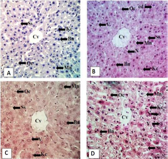
Effect of exposure of feed to microwaves in different contains on histological structure of liver of male mice. A: Control showing well organized mononucleated (mn) and bi nucleated (bn) hepatocytes of similar size in cord like structure having round nuclei. Sinusoidal spaces (Ss) of equal width lies between hepatic cords, round oval cells (Oc) observed in sinusoidal spaces. Central vein (Cv) is compact. Kupffer cells (Kc) elongated in shape lining the hepatocytes. B: Direct group represented decreased number of hepatocytes as well as nuclear vacuolation (V), necrosis (N) and cellular degeneration (cd). C: Glass group showed decreased number of hepatocytes (Bn and Mn) of variable size and shape, nuclear vacuolation and necrosis. Dilated central vein with infiltrated cells in its lumen. Sinusoidal spaces became wider. D: Plastic group also showed dilated sinusoidal spaces and central vein having infiltrated cells. Number of oval and Kupffer cells was decreased and nuclear vacuolation was also observed. Stain: hematoxylin and Eosin Magnification: 400X.

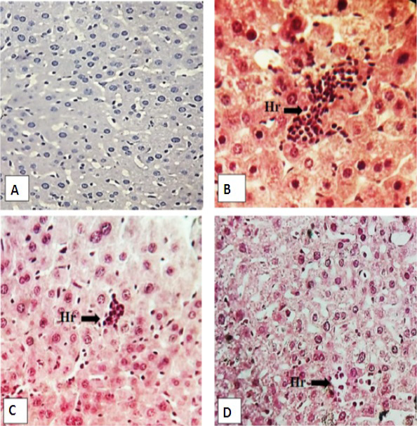
Effect of exposure of feed to microwaves in different contains on histological structure of male mice liver. A: Direct group showed hepatocytes regeneration (Hr) and disorganized structure of sinusoids (Ss) similarly glass. (B) and plastic group (C) also represented the hepatocytes regeneration zone and disorganized sinusoidal spaces. Stain: hematoxylin and Eosin Magnification: 400X.
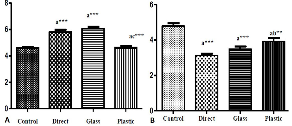
(A) Relative Nucleo-cytoplasmic index of mononuclear hepatocytes (µm). (B) Effect of microwave exposed feed on mean number of oval cells per unit area (10 cm2) in control and treated liver of adult mice after 28 days exposure of microwaves treated feed in different containers. Values are presented as Mean ± SEM, a = Control group vs treated groups, c = Glass group vs plastic group. ***P<0.001
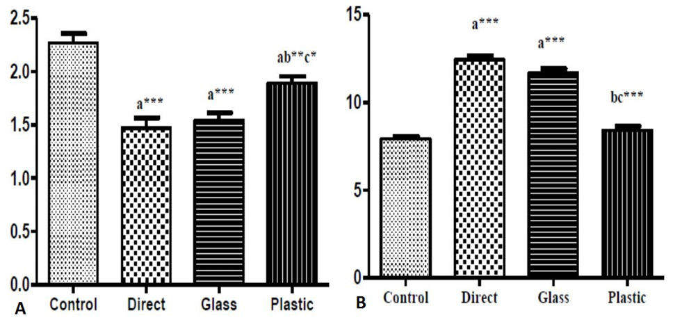
(A) Effect of microwaves exposed feed on mean number of Kupffer cells per unit area (10 cm2) in liver of control and treated mice after experimental exposure of 28 days. (B) Effect of microwaved exposed feed in different containers for four weeks on ACSA of central vein (µm)in liver of adult male mice. Values are presented as Mean ± SEM, a = Control group vs treated groups, b = Direct group vs glass and plastic groups and c = Glass group vs plastic group. ***P<0.001, **P<0.01, *P<0.05


