

Effects of Cypermethrin on the Hematological Parameters, Biochemical Components of Blood and Histopathological Changes in Different Organs of Chirruh Snow Trout (Schizothorax esocinus)


Effects of Cypermethrin on the Hematological Parameters, Biochemical Components of Blood and Histopathological Changes in Different Organs of Chirruh Snow Trout (Schizothorax esocinus)
Naveed Akhtar1*, Muhammad Fiaz Khan1*, Sadia Tabassum1,
Munawar Saleem Ahmad2 and Khan Dil Badshah3
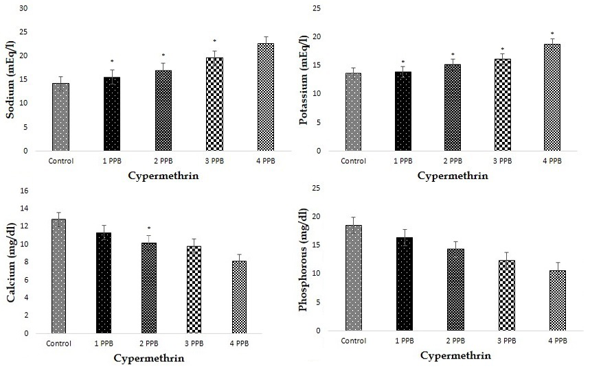
Effect of different concentration of cypermethrin on electrolytes level of S. esocinus.

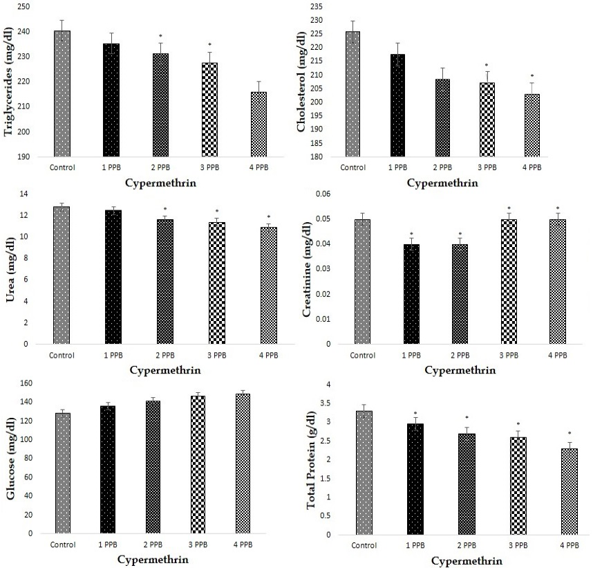
Effect of different concentration of cypermethrin on various biochemical components of S. esocinus.

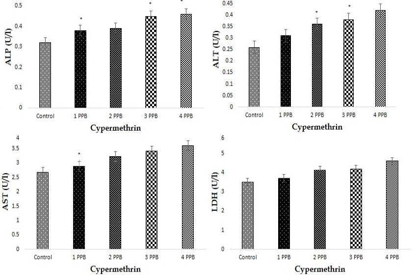
Effect of different concentration of cypermethrin on liver function enzymes in blood serum.
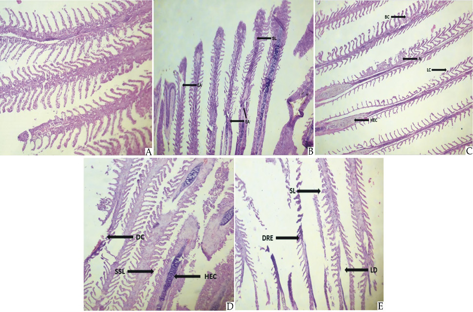
Effect of different concentration of cypermethrin on Gills of Schizothorax esocinus. (A) Control showing no lesions no necrosis and have normal primary and secondary lamellae. (B) fishes exposed to 1 ppb showing (SL) Shortening of lamellae and (EA) Erosion of gills arch. (C) fishes exposed to 2 ppb showing (BC) Blood congestion, (N) Necrosis, (LC) Lamellar curling and (HEC) Hypertrophy of epithelial cells. (D) fishes exposed to 3 ppb showing (DC) Degenerative changes, (SSL) Shortening of secondary gills lamellae and (HEC) Hypertrophy of epithelial cells. (E) fishes exposed to 4 ppb showing (SL) Shortening of lamellae, (DRE) Degeneration of epithelial cells and (LD) Lamellar destruction.

Effect of different concentration of cypermethrin on various histological structure of Brian of Schizothorax esocinus. (A) Control showing normal fish brain having no discolouration, no lesions, no morphological changes and have normal hippocampus. (B) fish exposed to 1 ppb showing (P) Pyknosis and (DC) Degenerative changes. (C) fish exposed to 2 ppb showing (P) Pyknosis, (N) Necrosis and (BC) Blood congestion. (D) fish exposed to 3 ppb showing (BC) Blood congestion, (N) Necrosis and (P) Pyknosis. (E) fish exposed to 4 ppb showing (BC) Blood congestion, (N) Necrosis and (P) Pyknosis.

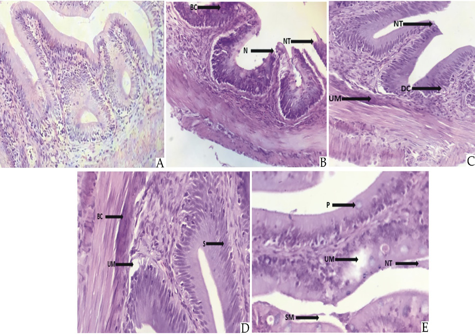
Effect of different concentration of cypermethrin on various histological structure of intestine of Schizothorax esocinus. (A) Control showing normal structure of intestine, epithelium, serosa and sub mucosa. (B) fish exposed to 1 ppb showing (BC) Blood congestion, (N) Necrosis and (NT) Necrosis of tip. (C) fish exposed to 2 ppb showing (NT) Necrosis of tip, (DC) Destructive changes and (UM) Ulceration of mucosa. (D) fish exposed to 3 ppb showing (BC) Blood congestion, (S) structural integrity loss and (UM) Ulceration of mucosa. (E) fish exposed to 4 ppb showing (UM) Ulceration of mucosa, (P) Pyknosis, (NT) Necrosis of tip and (SM) Shrinkage of mucosa.

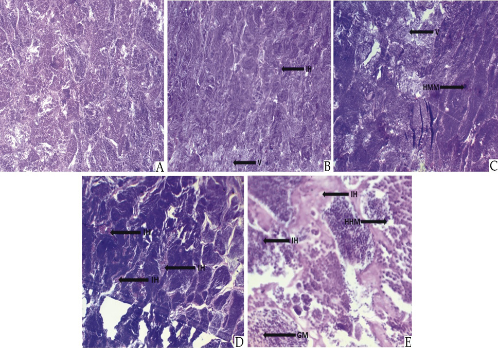
Effect of different concentration of cypermethrin on various histological structure of Kidney of Schizothorax esocinus. (A) Control showing histology of kidney with normal glomerulus, tubules and interstitial tissues. (B) fish exposed to 1 ppm showing (V) Vacuolation and (IH) Interstitial hemorrhage. (C) fish exposed to 2 ppm showing (HMM) Hyperactivated MM and (V) Vacuolation. (D) fish exposed to 3 ppm showing (IH) Interstitial hemorrhage. (E) fish exposed to 4 ppm showing (IH) Interstitial hemorrhage, (GM) Multifocal granulomas and (HMM) Hyperactivated MM.
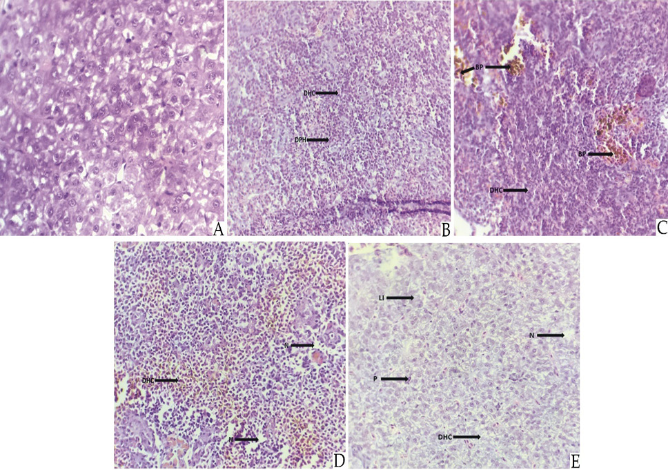
Effect of different concentration of cypermethrin on various histological structure of Liver of Schizothorax esocinus. (A) Control showing normal hepatocytes, hepatopancrease and sinusoids. (B) fish exposed to 1 ppb showing (DHC) Disappearance of hepatic cell wall and (DPH) Densly packed hepatocytes. (C) fish exposed to 2 ppb showing (BP) Bile pigment and (DHC) Disappearance of hepatic cell wall. (D) fish exposed to 3 ppb showing (DHC) Disappearance of hepatic cell wall and (N) Necrosis. (E) fish exposed to 4 ppb showing (Li) Leukocyte infiltration, (DHC) Disappearance of hepatic cell wall, (P) Pyknosis and (N) Necrosis.

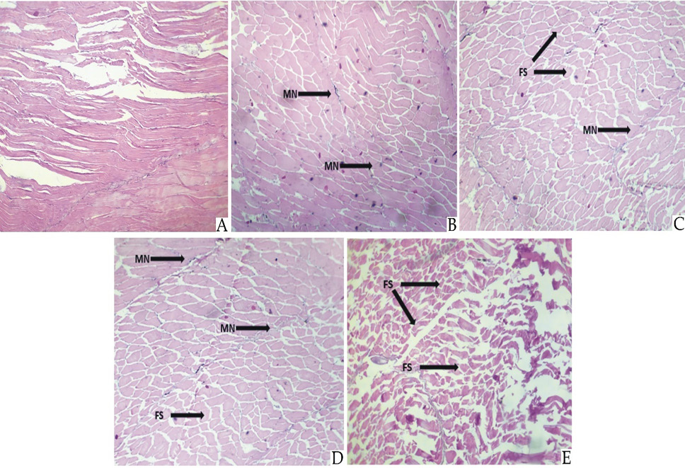
Effect of different concentration of cypermethrin on various histological structure of smooth muscles of Schizothorax esocinus. (A) Control showing normal fish muscle having no necrosis and fragmentation. (B) fish exposed to 1 ppm showing (MN) Muscles necrosis. (C) fish exposed to 2 ppm showing (FS) Fragmentation of sarcoplasm and (MN) Muscles necrosis. (D) fish exposed to 3 ppm showing (MN) Muscles necrosis and (FS) Fragmentation of sarcoplasm. (E) fish exposed to 4 ppm showing (FS) Fragmentation of sarcoplasm.

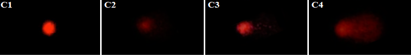
Effect of different concentration of cypermethrin on the DNA damage in peripheral blood erythrocytes using comet assay in control and treated group of Schizothorax esocinus. C1, I ppb; C2, 2 ppb; C3, 3 ppb; C4, 4 ppb.

