
Evaluation of Hemato-Biochemical Parameters, Histopathological Altration and Antioxidant Trace Elements in Demodicosis Infected Dogs


Evaluation of Hemato-Biochemical Parameters, Histopathological Altration and Antioxidant Trace Elements in Demodicosis Infected Dogs
Noha M El-Motaily1, Ossama M Abdou1, Heba S Farag1, Kawkab A Ahmed2, Mahmoud Saber1*

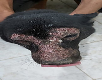
Figure 1:
Dog showing localized demodicosis.

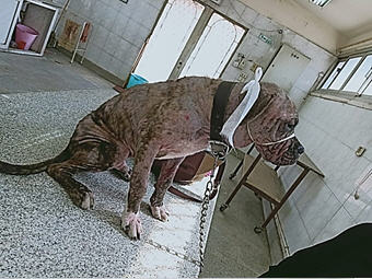
Figure 2:
Dog showing generalized demodicosis.

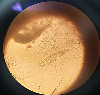
Figure 3:
Demodex Canis adult mite under microscope.

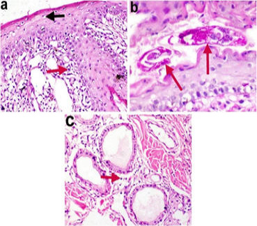
Figure 4:
Photomicrographs of a skin biopsy of generalized demodicosis infected dog showing (a) vacuolar degeneration of the epidermal prickle cells (black arrow) and severe dermatitis (red arrow). (b) Demodex mite in the follicle (red arrows). (c) periglandular inflammatory cells infiltration (red arrow) (H & E, X 200 (a & c ), X 400 (b).

July 2022
Vol. 10, Iss. 7, Pages 1423-1658
