
Histopathological Studies of Pond Reared Indian Major Carp, Catla catla Infested with Argulus japonicus and Trial for Argulosis Treatment


Histopathological Studies of Pond Reared Indian Major Carp, Catla catla Infested with Argulus japonicus and Trial for Argulosis Treatment
Md. Abdullah Al Mamun1,2*, Shamima Nasren1,3, Sanjay Singh Rathore1 and Kavalagiriyanahalli Srinivasiah Ramesh1
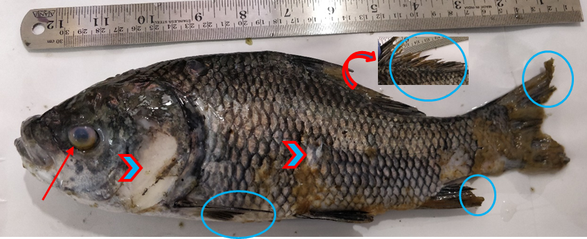
Argulosis in catla showing numerous gross lesions including fin rots (circles), skin and scal loss (arrow heads), corneal opacity (arrow), excessive mucus on the surface of the infested fish.
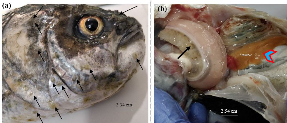
Heavy infestation of Argulus japonicus (a) A. japonicus firmly attached on the operculam and head surface of catla (arrows); (b) Infested catla showed pale gills and clogged gill arch (arrow) with englarge liver that lost its original reddish-brown colour and turned to yellowish-brown colour (arrow heads).

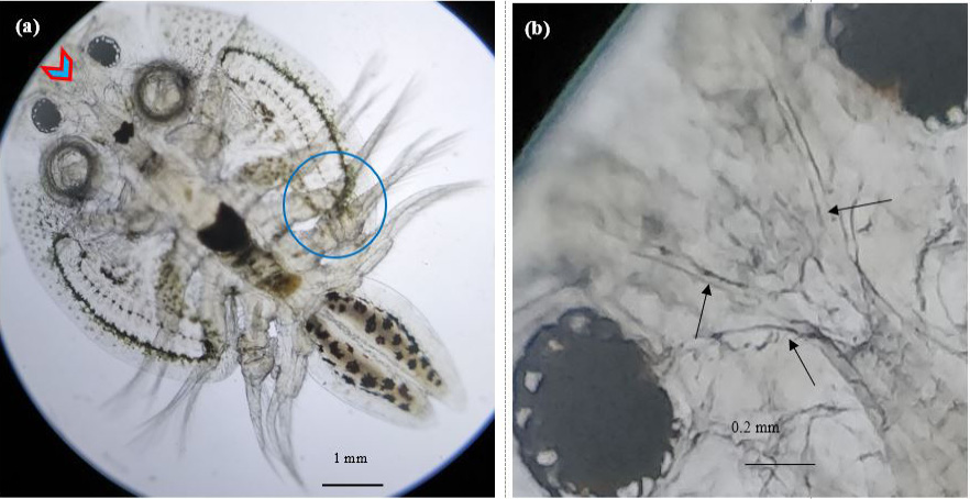
(a) Argulus japonicus (Thele 1900) with distinct morphological features including branched dorsal ridge (arrow head) and bilobed second segment (circle); (b) Computer blown up image showing clear image of branched dorsal rigdge (arrows).
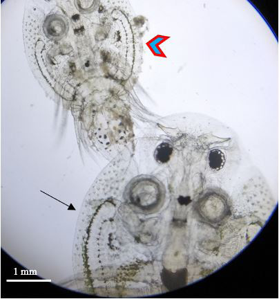
Juvenile (arrow head) and adult (arrow) Argulus japoncus found in catla.

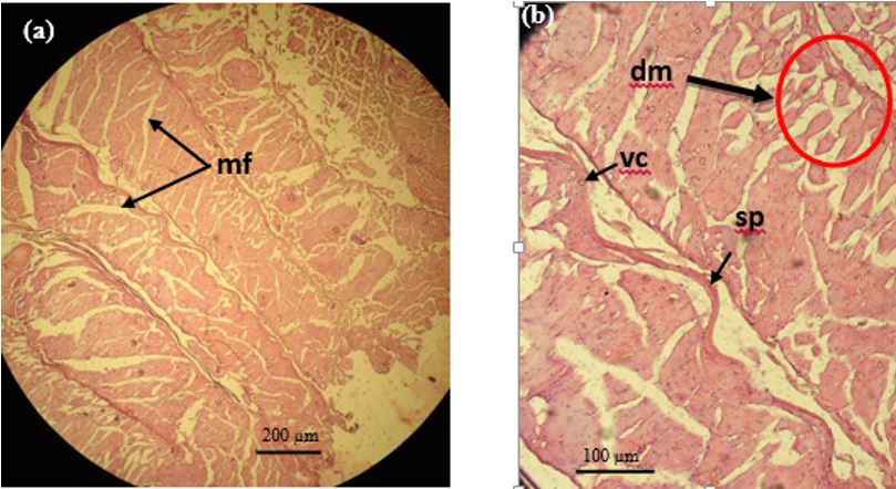
Photomicrographs of the muscle of catla infested with Argulus japonicus. a) The broken of myofibrils (mf) (10×); b) Splitting of muscle fibres (sp) disintegrated myotomes (dm) and vacuolation (vc) within the myotomes (40×).

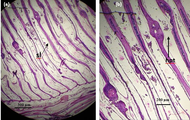
Gill histopathology of catla infected by Argulus japonicus a) Displaying complete loss of secondary gill lamellae (sl) and irregular shaped primary gill lamellae (10×); b) Part of the primary gill lamellae become hypertrophied (hpt) (40×).

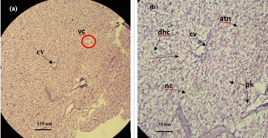
Liver histological section of infested catla by Argulus japonicus, a) Central vein (cv) vacuolization (vc) (10×); b) Numerous pyknotic nuclei (pk), disintegrated hepatocyte cells (dhc), atrophied nucleus (atn), necrotic hepatocytes (nc) (40×).
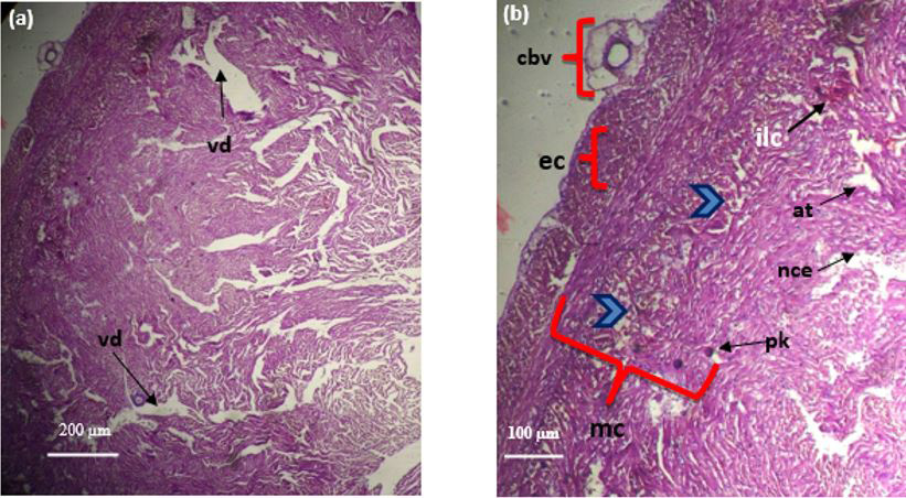
Photomicrograph of heart ventricle of catla infested with Argulus japonicus. a) Vacuolar degeneration (vd) accompanied with disrupted myocardium tissues (10×); b) Coronary blood vessel (cbv), epicardium (ec) myocardium (mc), cardiac muscle showed necrotizing endocardium (nce), infiltrated lymphocytes (ilc), degenerated myocardium (arrow heads) accompanied with atrophy (at) and hypertrophied cells (pk) (40×).

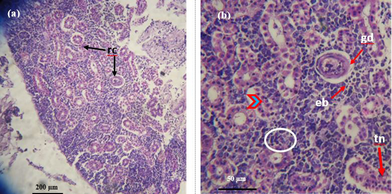
Photomicrographs of the infested kidney of catla by Argulus japonicus. a) Showing renal corpuscles (rc) (10×); b) Glomerular distension (gd) accompanied with extended bowmans capsule (eb), vacuolation (arrow head) tubular necrosis (tn), increase in the presence of leukocyte cells (circle) (40×).

Histology section of fore gut of catla infested with A. japonicus. a) Normal architecture of villi (v) with numerous gut associate lymphoid tissues in mucosal tissues (red circle) (10×); b) Emptied mucous cells (mc), aggregated lymphocyte cells (lc) (40×) =artifacts.
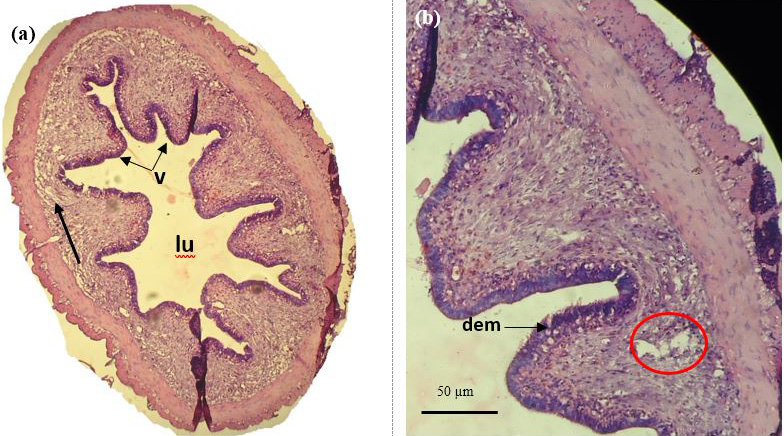
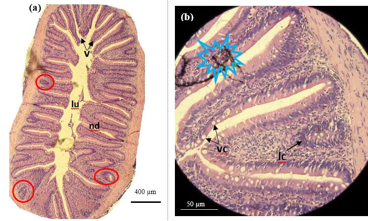
Histological analysis of mid gut of catla infested with Argulus japonicus. a) Transvers section showing short and stout villi (v) large lumen space (lu) with lamina propria (lp) disruption of mucosal and sub-mucosal tissues (arrow) (10×) b) Damage of enterocytes and microvilli (dem) with large vacuolar space (circle) (40×).
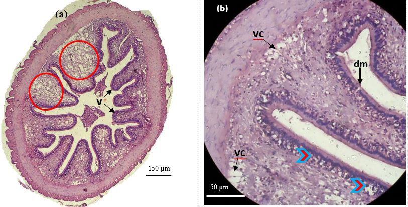
Histology section of hind gut of catla infested with Argulus japonicus. a) Short and stout villi (v), disruption and vacuolization (vc) in the mucosal tissues (red circle) (10×); b) disintegration of enterocytes (arrow heads), damage of microvilli (dm) (40×).
