

Isolation, Cultivation and Identification of Spermatogonial Stem Cells from Juvenile Buffalo Testes


Isolation, Cultivation and Identification of Spermatogonial Stem Cells from Juvenile Buffalo Testes
Huan Yang1, Tingting Li1, Huimin Zhao1,2, Xiaoyuan Zhang1, Huiyan Xu1, Yangqing Lu1, Xingwei Liang1, Shengsheng Lu1, Xiaogan Yang1*and Kehuan Lu1*
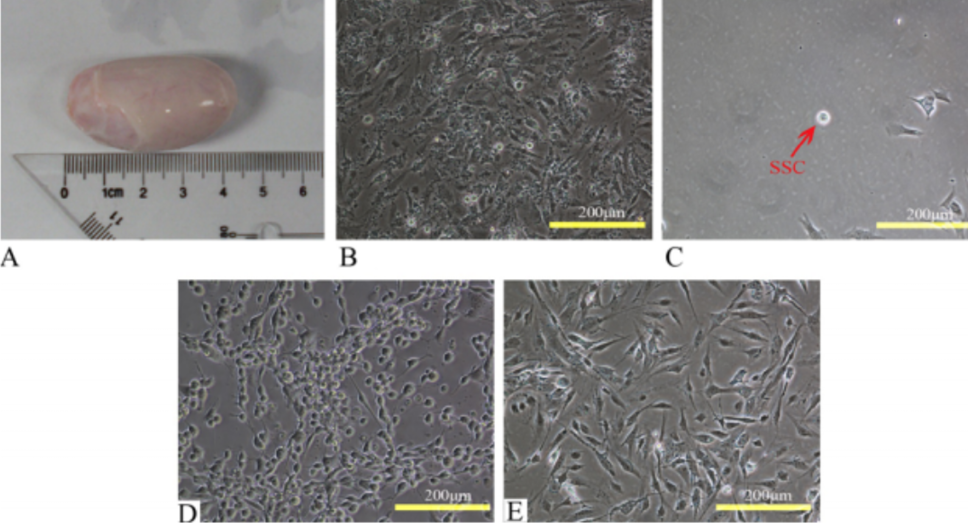
Differential adherence enrichment of buffalo SSCs. A, Testes from a 3 month-old buffalo; B, in gelatin-treated culture dishes, testicular somatic cells adhere to the surface of the dish before germ cells, and germ cells do not adhere firmly; C, when purified in a culture dish coated with rat tail collagen, the adherent cells mainly consist of spermatogonia; D, co-culture of separated germ cells with STO; E, separated testicular somatic cells (mainly Sertoli cells).
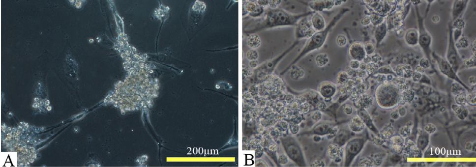
In vitro culture of buffalo SSCs. SSCs attached to STO feeder layer for growth. A, The isolated and cultured buffalo SSCs were found in typical grape clusters; B, the frozen-thawed buffalo SSCs had no change in morphology and performance.
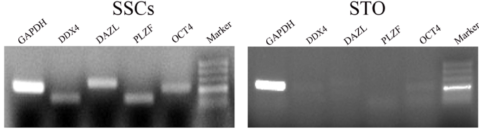
Gene expression of buffalo SSCs and STO. DDX4 and DAZL are germline-related markers, PLZF is a buffalo spermatogonia marker and OCT4 is a pluripotency marker.
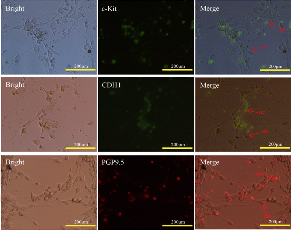
Cellular immunochemical staining of buffalo SSCs. Grape-like cluster of SSCs expressing stem cell factors such as c-Kit, undifferentiated spermatogonia marker PGP9.5 and CDH1.
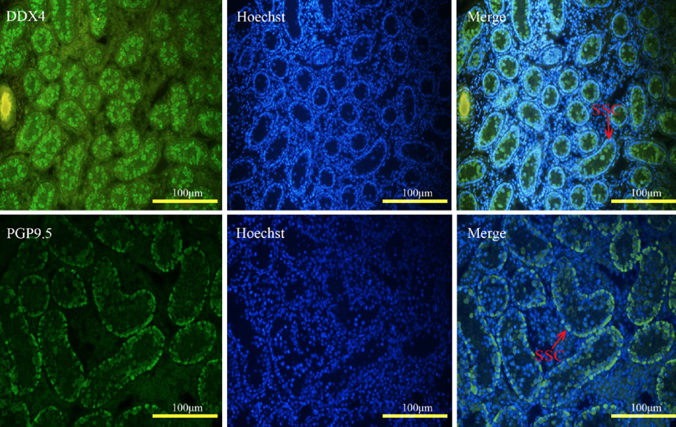
Expression of the germline marker DDX4 and undifferentiated spermatogonia marker PGP9.5 in buffalo testes tissue.

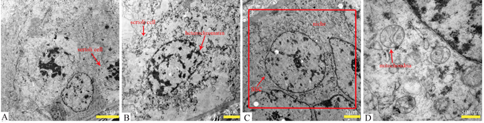
Sertoli cells and niche in seminiferous tubules of buffalo testes under transmission electron microscopy. A-B, The shape of Sertoli cells is irregular, and heterochromatin is abundant; C, the SSCs are surrounded by Sertoli cells, which form a niche structure; D, the SSCs in testes have spherical mitochondria and few mitochondrial cristae.
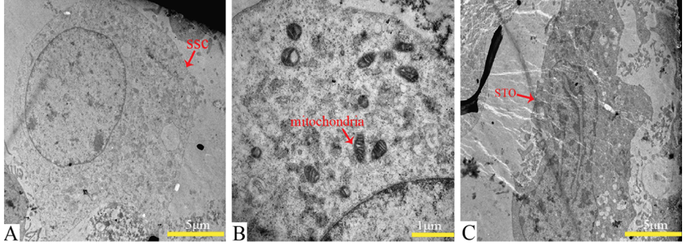
In vitro cultured SSCs and STO under transmission electron microscopy. A, SSCs have a high nucleus-cytoplasm ratio, are 15 μm in diameter, and have less heterochromatin; B, SSCs cultured in vitro have more mitochondrial cristae; C, apoptotic STO cell.

