

Light and Scanning Electron Microscopic Studies on Eustrongylides exciscus Larvae (Nematoda: Dioctophmida) from Channa punctatus Bloch from India


Light and Scanning Electron Microscopic Studies on Eustrongylides exciscus Larvae (Nematoda: Dioctophmida) from Channa punctatus Bloch from India
Neelima Gupta

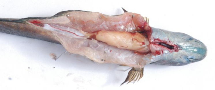
L4 dissected Channa punctatus showing Eustrongyloides tubifex lying in the body.
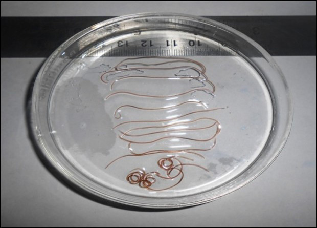
L4 Eustrongylides tubifex isolated in a petri dish from a single fish.
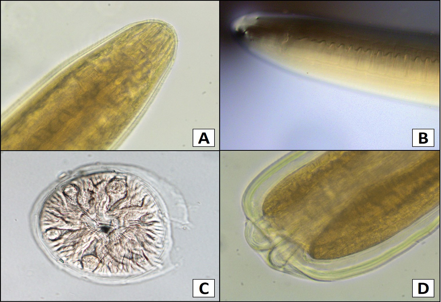
Light microphotographs of L4 Eustrongylides tubifex: A, anterior end showing tapering end; B, anterior end showing papillae under fine adjustment; C, en face view showing arrangement of papillae; D, posterior blunt end.

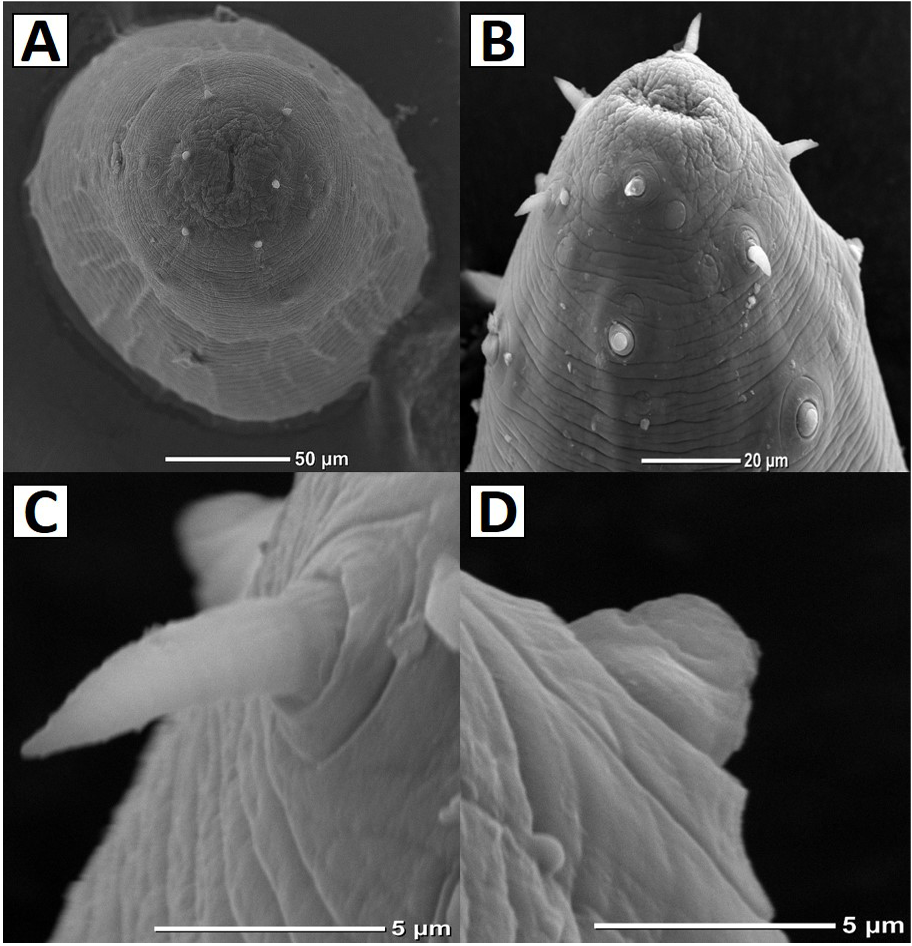
Scanning electron micrographs of L4 Eustrongylides tubifex: A, en face view showing slit-like mouth (arrow) and number of papillae; B, anterior end showing arrangement of papillae; C, inner papilla showing narrow base and spine-like apex; D, outer papilla having wide base and nipple-like apex.

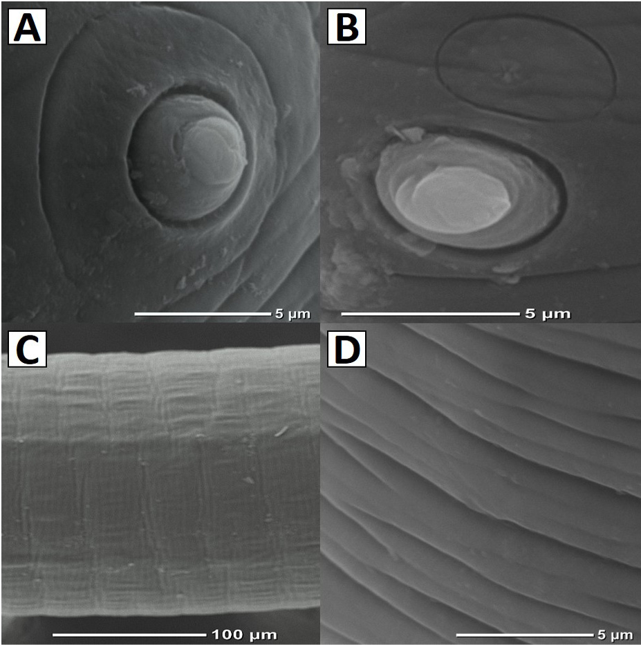
Scanning electron micrographs of L4 Eustrongylides tubifex: A, surface view of outer papilla encircled in a ring; B, sensillae situated above the outer papilla; C, body showing striations; D, striations under higher magnification (note the grooves and incomplete margins).


