

Marsh Frog (Pelophylax ridibundus) as a Bioindicator to Assess Pollution in an Agricultural Area


Marsh Frog (Pelophylax ridibundus) as a Bioindicator to Assess Pollution in an Agricultural Area
Turgay Şişman1,*, Muhammet Çağrı Keskin1, Hatice Dane1, Şeymanur Adil1, Fatime Geyikoğlu1, Suat Çolak2 and Esra Canpolat1

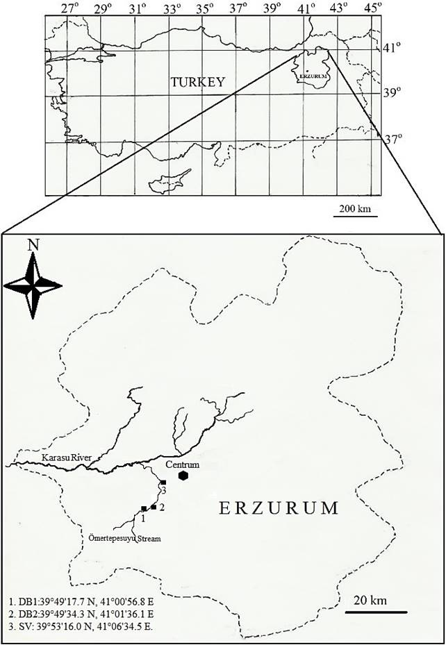
The sampling stations.

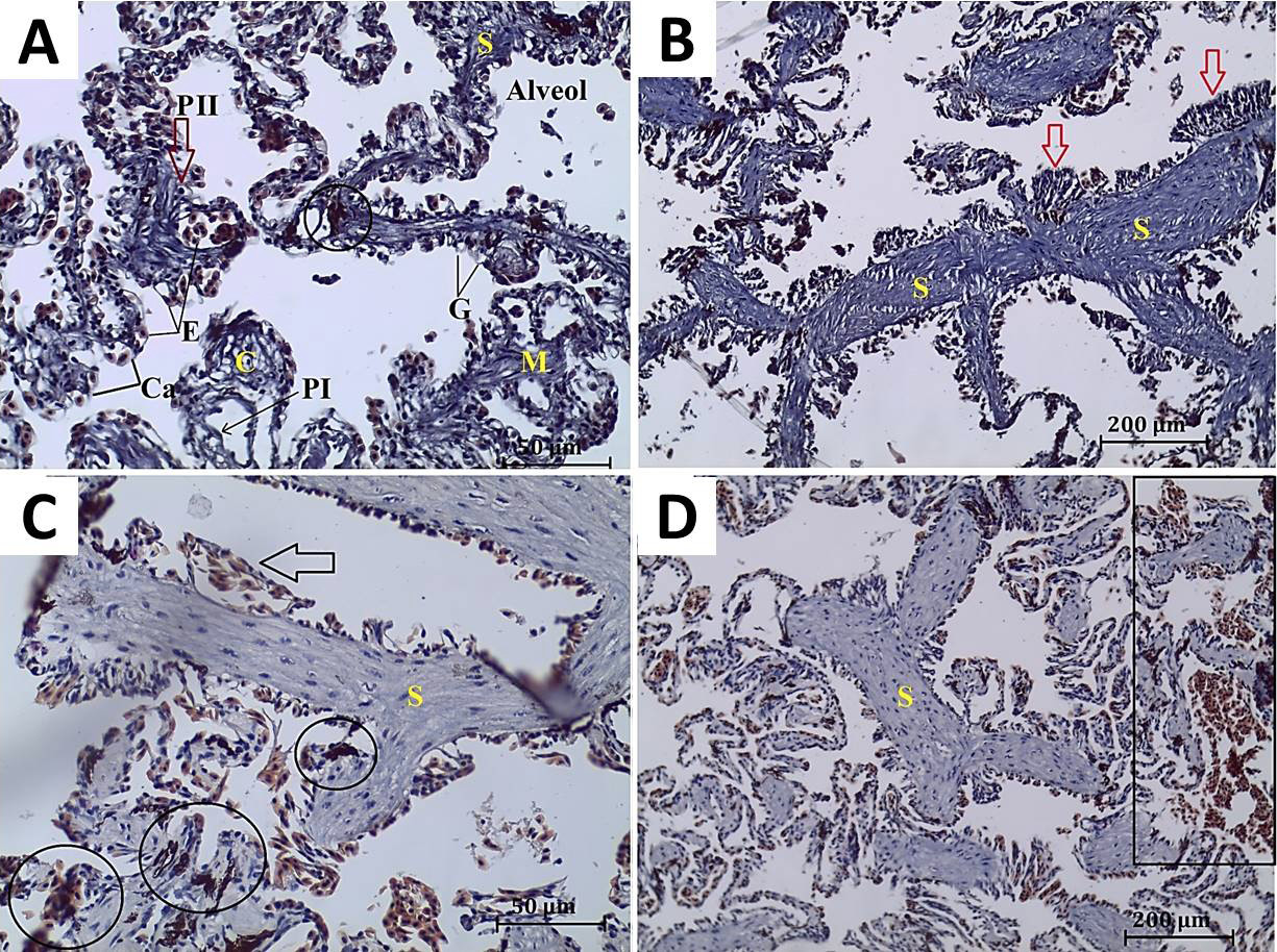
A, normal alveolar construction of P. ridibundus collected from DB1. Septum (S), type I and II pneumocytes (PI, PII), erythrocytes (E), goblet cells (G), capillaries (Ca), alveol, melanomacrophage aggregate (encircled), connective (C) and muscle tissue (M). B, C and D, histopathological alterations of the lung of P. ridibundus: B: Samples of DB2; C, D: Samples of SV. B, thickness of alveolar septum and hyperplasia of alveolar epithelium (arrows). C, dilated blood capillaries (arrow) and melanomacrophage aggregation (encircled). D, congestion (squared), (H&E).
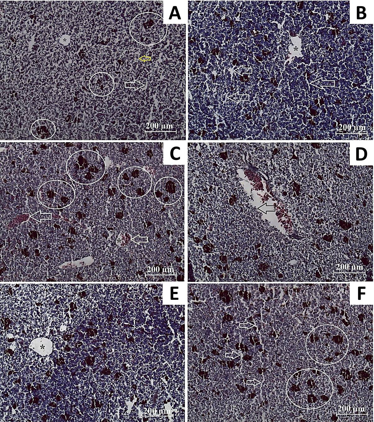
Histological structures and alterations of the liver of P. ridibundus. A; samples of DB1, B, C; samples of DB2: A, sinusoids (white arrow), central vein (asterisk), hepatocytes (yellow arrow) and melanomacrophage aggregation (encircled). B, degeneration of central vein (asterisk) and sinusoidal dilatation (arrows). C, increasing melanomacrophage aggregation (encircled), congestions of the central (asterisk) and portal vein (arrows). D, E, F samples of SV. D, degeneration of vascular epithelium (arrow). E, non-homogenous parenchyma. F, sinusoidal dilatations (arrows) and increasing melanomacrophage aggregation (encircled), (H&E).
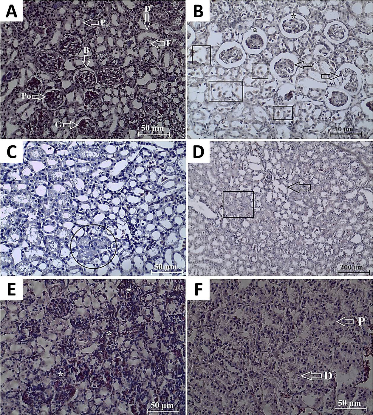
A, normal histological properties of the kidneys of P. ridibundus from DB1: renal corpuscles with glomerulus (G) and Bowman’s space (B); podocyte (Po), erythrocytes (E), proximal (P) and distal tubules (D). B, C and D; histological alterations of the specimens of DB2. B, glomerulonephritis (black arrows), congestion in renal parenchyma (squared), and expansion of Bowman’s space (asterisks). C, tubular necrosis (encircled). D, tubular dilatation (arrow), tubule lumens as eosinophilic in appereance (squared). E, F; Histological changes in the kidney of SV specimens. E, lymphocytes infiltration (asterisks). F, degeneration of proximal (P) and distal (D) tubules, (H&E).

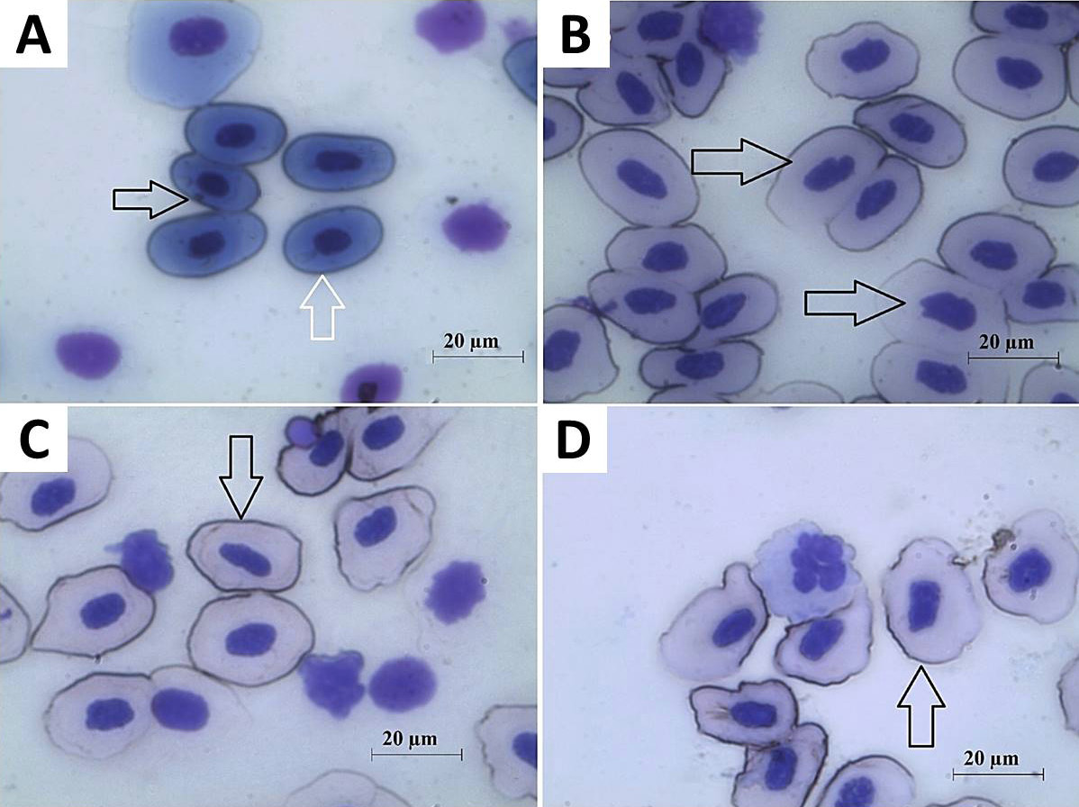
The ENAs recorded in P. ridibundus: A, Normal (white arrow) and micronucleus (black arrow); B, notched nucleus (black arrows); C, kidney-shaped nucleus (black arrow); D, lobed nucleus (black arrow), Giemsa.


