
Molecular Detection of FMDV Serotype A Isolated from the Egyptian Delta During 2019-2020


Molecular Detection of FMDV Serotype A Isolated from the Egyptian Delta During 2019-2020
Gawhara Ahmed-Abdelmonem1*, Zeinab Aboezz2, Ahmed Habashi1, Saad Sharawi2

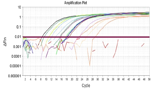
The amplification plots of Pan-serotypic (3D) qRT-PCR assay of collected field samples.

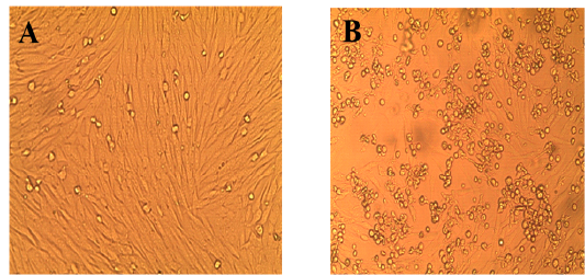
Pictures showing the FMDV’s cytopathogenic effect on inoculated BHK-21 cells. Panel (A) control BHK 21 cell line, Panel. (B) BHK21 Cells showing characteristic CPE.

Serotyping of FMDV in 10 oral epithelial samples by conventional RT-PCR.
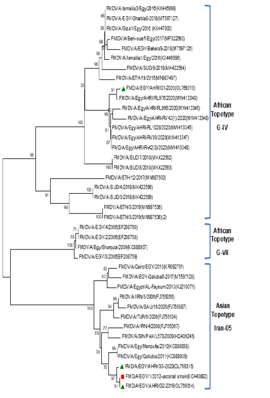
Phylogenetic tree constructed using the neighbor joining method depending on the VP1 gene sequences of the three FMDV isolates identified in this study and 36 additional FMDV serotype A sequences obtained via the GenBank (https://www.ncbi.nlm.nih.gov/genbank) dataset. The bootstrap probabilities are signified by numbers at the internal nodes (1000 replicates). Red square refers to vaccinal strain, green triangles refer to isolates of present study.

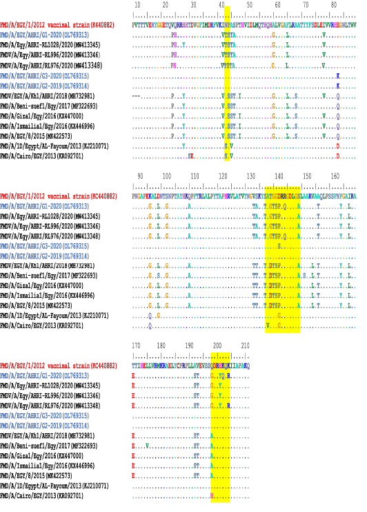
Deduced amino acid sequences within the VP1 gene of the new FMDV isolates in this study were compared to existing FMDV type A isolates over the years. The highlighted regions refer to the significant variable antigenic sites chosen in the comparison.

