

Molecular Detection of Hepatitis E Virus in Tibetan Swine


Molecular Detection of Hepatitis E Virus in Tibetan Swine
Gong Ga1,2 and SuoLang Sizhu2,*

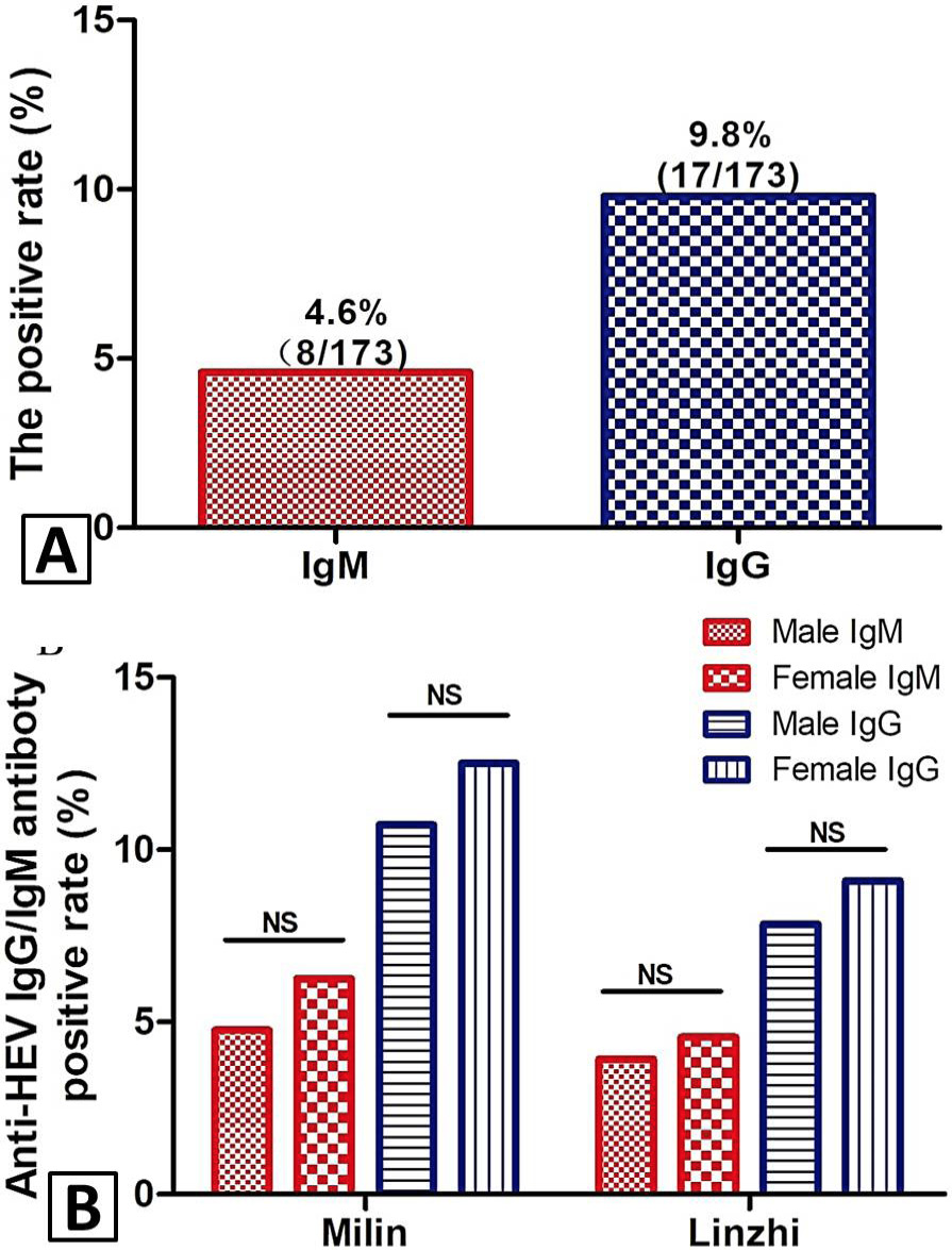
Seroprevalence of HEV in Tibetan swine. A, the positive rate of anti-HEV IgM and IgG antibodies in serum of Tibetan swine, China; B, the different of anti-HEV IgM antibody and anti-HEV IgG antibody positive rate between the gender in two cities; C, the different of anti-HEV IgG antibody positive rate between the gender in two cities.
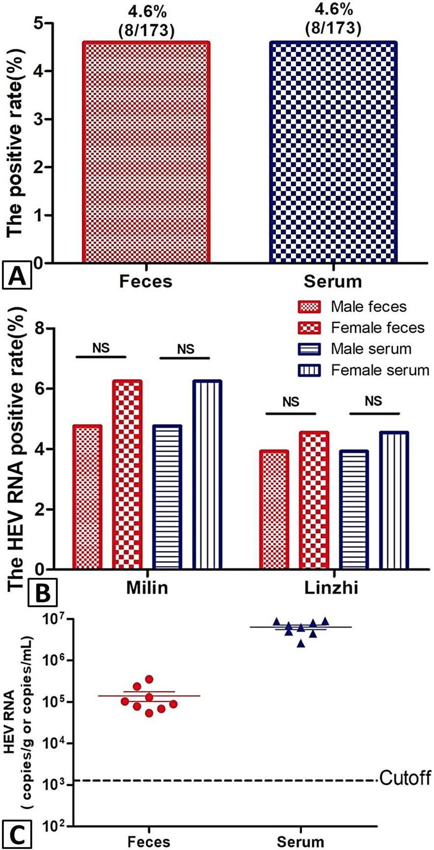
Prevalence of HEV infection in Tibetan swine. A, the positive rate of HEV RNA in feces and serum samples of Tibetan swine, China; B, the different of HEV RNA positive rate in feces and serum between the gender in two cities; C, the virus titer in feces and serum of Tibetan pigs, China. Unit for feces samples, copies/g; Unit for serum samples, copies/ml.
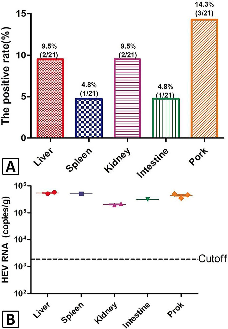
HEV infection in swine tissues. A, the positive rate of HEV RNA in tissue of Tibetan swine, China. B, the virus titer in tissue of Tibetan pigs, China. Unit for tissue samples, copies/g.

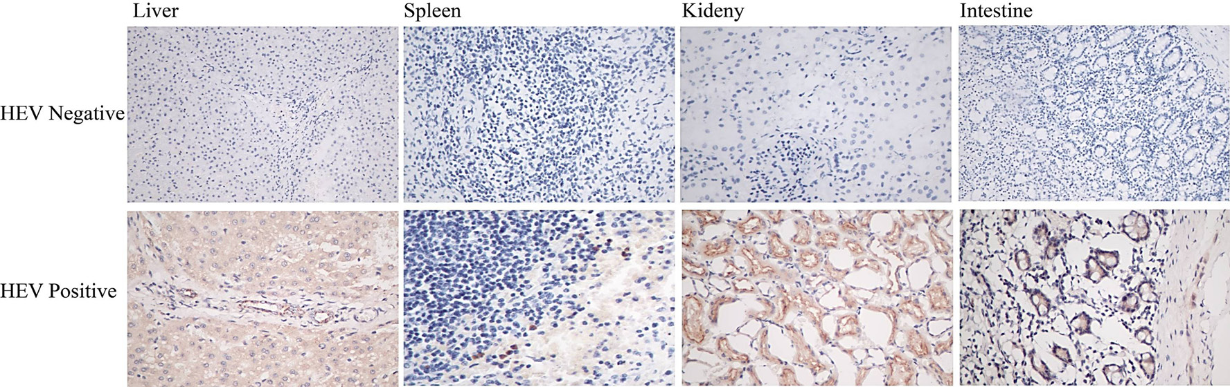
Immunohistochemical analyses of swine tissue. Immunohistochemical analyses (IHC) for HEV antigens in tissue samples of Tibetan swine (400×), including liver, spleen, intestine and kidney.


