
Pathological Changes Caused by Nematode Parasites in the Intestine of Domestic Pigeon (Columba livia) of Karachi, Sindh, Pakistan

Pathological Changes Caused by Nematode Parasites in the Intestine of Domestic Pigeon (Columba livia) of Karachi, Sindh, Pakistan
Rizwana Abdul Ghaffar* and Kamil Nadeem
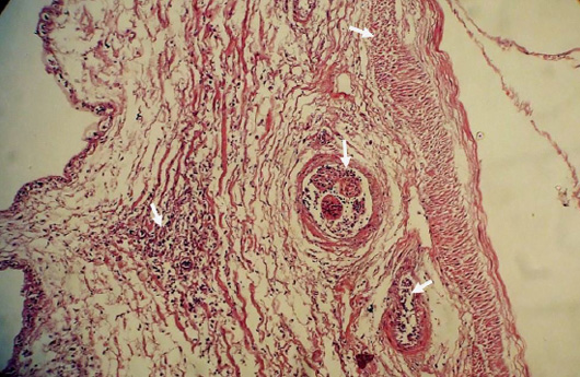
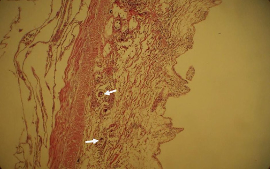
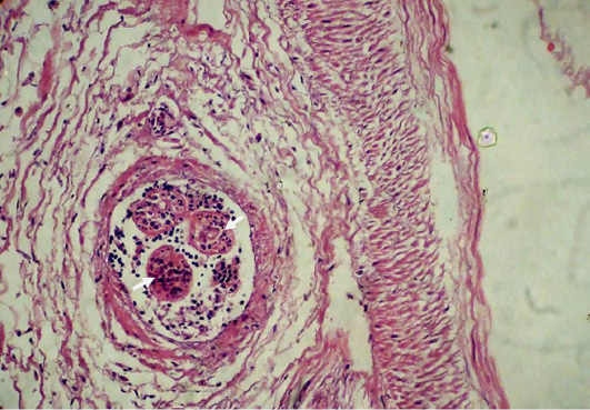
High magnification shows massive degenerative changes and infiltration of inflammatory cells (Arrows). Blood vessels appeared with a thick fibrotic wall. Migratory tunnels and penetration sites of parasites are also obvious. × 200.

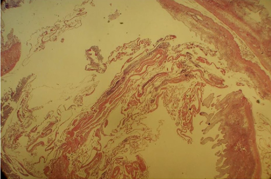
Section of intestine infected with nematode (Ascaridia sp. and Capillaria sp.) shows atrophy and fibrosis of mucosa and sub-mucosa. × 40.

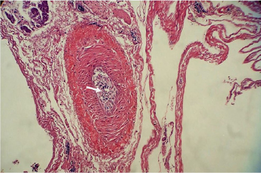
A section of the intestine infected with nematodes shows the complete destruction of all cellular layers. Hypertrophy of villous crypt is observed whereas villi are completely washed out. Fibrosis of mucosa and sub-mucosa is also obvious. Section of nematode larvae (Ascaridia sp.) observed in this section. (Arrow) × 100.
Photomicrograph of the intestine section infected with nematodes (Ascaridia sp. and Capillaria sp.) shows total dystrophy of tissues. Aggregation of eosinophils (arrow) is obvious among necrotic tissue. × 40.

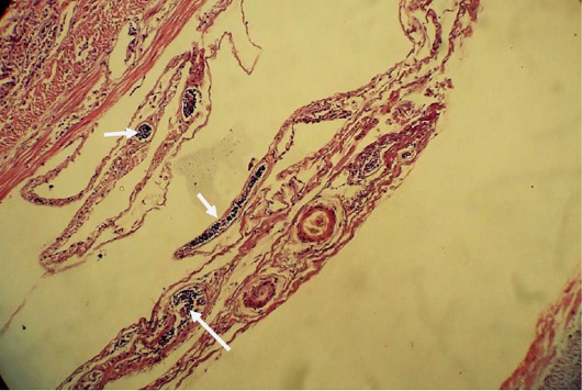
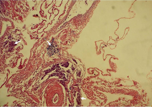
A portion of the intestine shows migratory tunnels formed due to nematodes (Ascaridia sp.) in muscularis mucosa and sub-mucosa. Aggregation of eosinophils found at the infestation site of parasites (arrow). Infiltration of inflammatory cells is also obvious throughout the area. × 200.

High magnification shows proliferated blood vessels with inflammatory cells. (Arrow). × 400
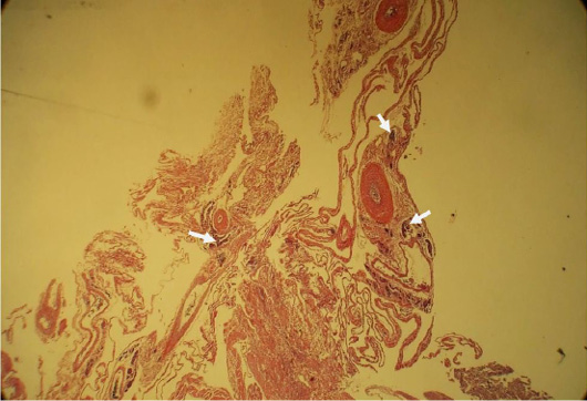
The micrograph of the intestine shows the orientation of cellular organization disintegrated by fibrotic infiltration. Eosinophils are scattered throughout the section due to heavy infection of cestodes and nematodes (Ascaridia sp. and Capillaria sp.) (Arrow) × 200.

High magnification shows degeneration of all layers. Villi are also completely washed out. Section of nematode larvae (Ascaridia sp.) surrounded by necrotic tissues. (Arrow) x400.
