

Phenotyping of Amphistomes, and Pathological, Hematological and Bile Biochemical Response to Gigantocotyle explanatum Infection in Buffaloes


Phenotyping of Amphistomes, and Pathological, Hematological and Bile Biochemical Response to Gigantocotyle explanatum Infection in Buffaloes
Sadaf Ijaz Malik1, Kiran Afshan2* and Mazhar Qayyum1
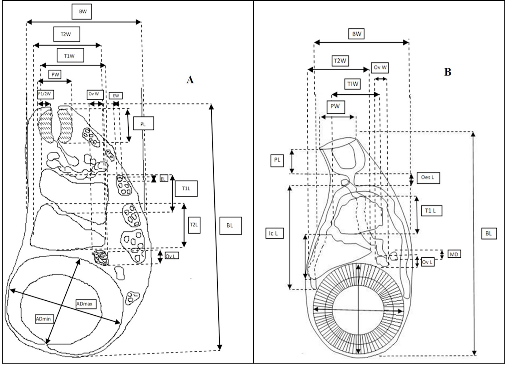
Standardized measurements of (A) Whole mount (B) Sagittal section of adult Amphistomes. The key to abbreviations for all measurements is shown in Table I.
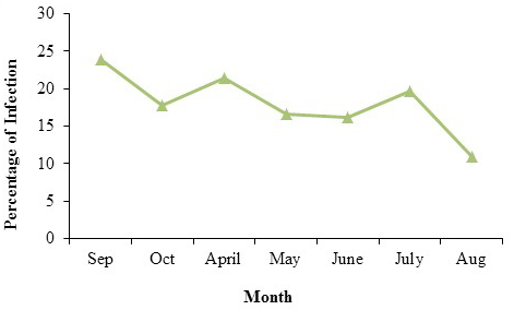
Prevalence of amphistomes in buffaloes of central Punjab in different months of the year 2010-2011.

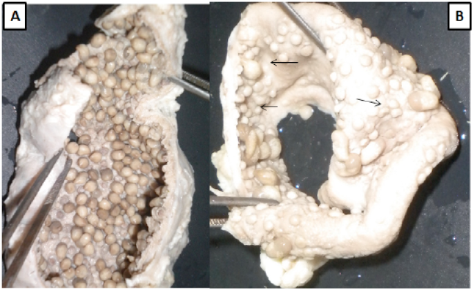
A bile duct contains numerous amphistomes (A) and dome-like protuberances” after removal of the amphistomes (B) indicated with arrows.

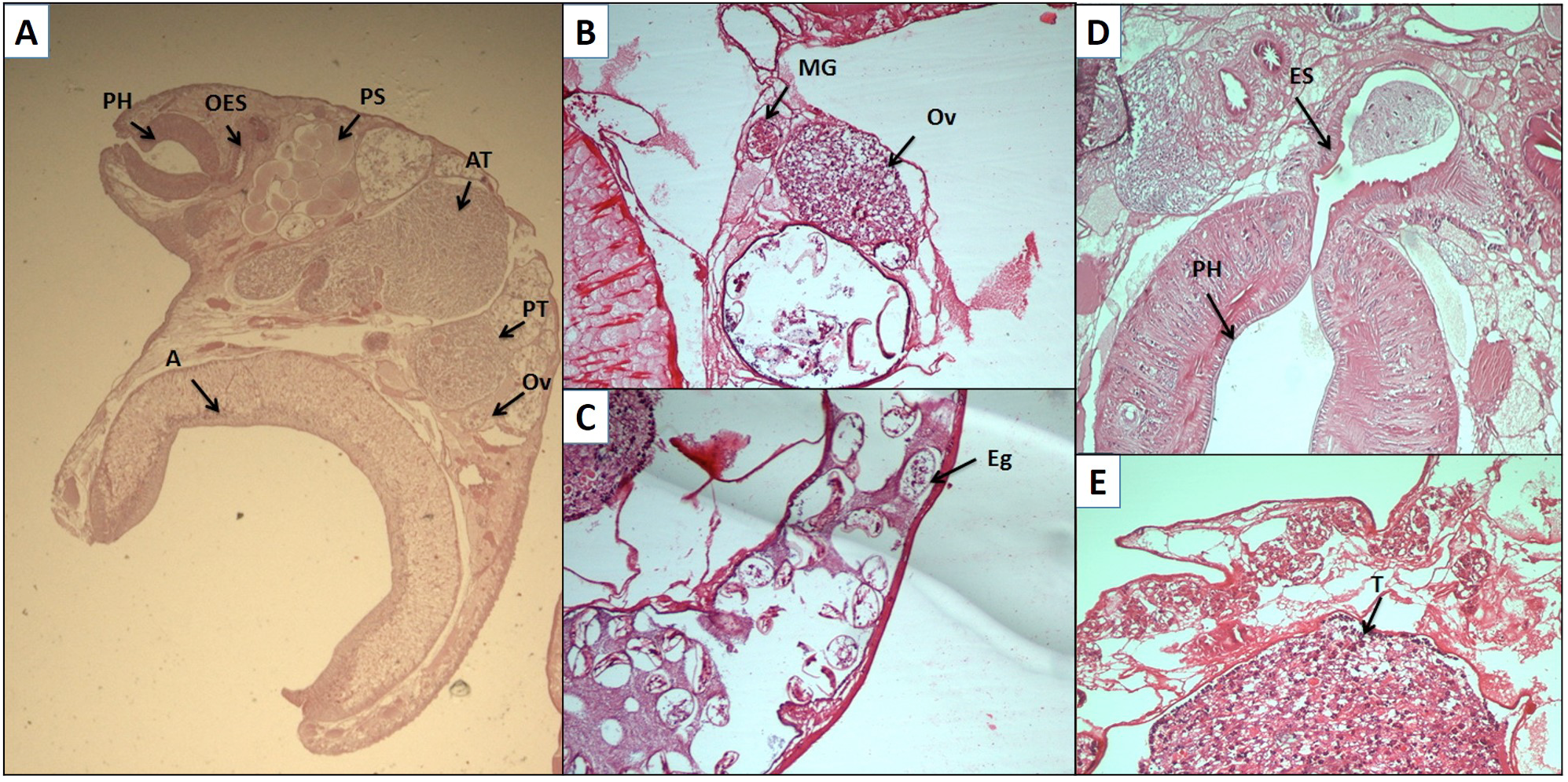
A, Representative sagittal sections of Gigantocotyle explanatum indicates PH, pharynx; Es, esophagus; PS, pars seminalis; AT, anterior testis; PT, posterior testis; Ov, ovary and A, acetabulum. B, an enlarged image of ovary and Mehli’s gland; C, egg; D, pharynx, esophagus; E, testis.
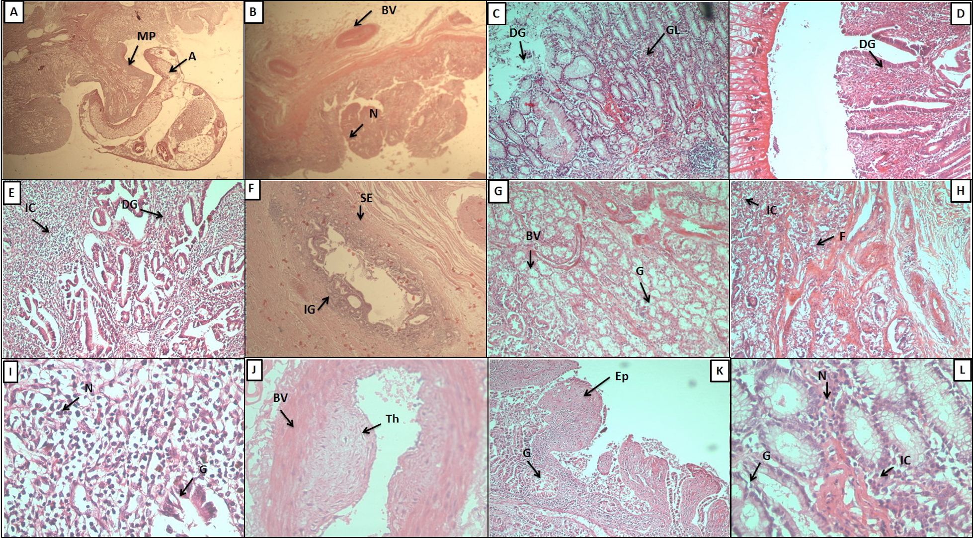
Histopathological study of buffalo bile duct. A: Cross section of the infected bile duct; A, acetabula; MP, mucosal plug. B: mucosal plugs formed due to parasite attachment; BV, blood vessel; N, nodule. C: the glandular mucosa showing extensive glandular hyperplasia; DG, degenerated gland; GC, glandular cells. D: the glandular degeneration and villi like appearance at the site of fluke attachment. E: massive glandular degeneration and inflammation; IC, inflammatory cells. F: glandular infiltration in serosal layer; IG, inflammed glands; SE, serosa. G: sub mucosal layer showing increased glandular hyperplasia; BV, blood vessel; G, gland. H: serosal layer of infected bile duct showing fibrosis; IC, inflammatory cells; F, fibrosis. I: inflammatory cells; N, nucleus; G: gland. J: thick walled blood vessel showing hyperplasia and thrombus formation. K: villi like appearance of hyperplastic mucosa and degeneration of upper epithelial layer; EP, epithelial layers; GL: gland. L: increased nuclear size in infected tissue; N, nucleus; IC, intestinal caeca.

