
Phylogenetic and Histopathological Characterization of Newcastle Disease Virus (VII.1.1) Recently Isolated from Naturally Infected Quails in Egypt


Phylogenetic and Histopathological Characterization of Newcastle Disease Virus (VII.1.1) Recently Isolated from Naturally Infected Quails in Egypt
Mohamed Lebdah1, Sahar Abd El Rahman2*, Ahmed Attia3, Reham Karam2, Naglaa Fathy Saeed Awad1, Mohamed Ibrahim El Bagoury1
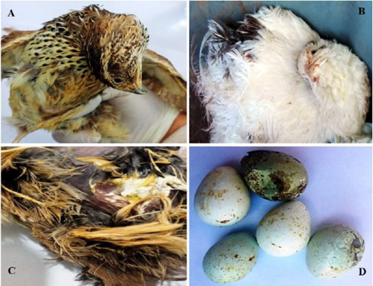
Clinical signs in naturally infected quails 2 months age from Egypt: (A, B) Nervous signs including torticollis and opisthotonos; (C) Digestive disorder including whitish and greenish diarrhea; (D) Deterioration of egg including broken, thin shell and loss of pigmentation.

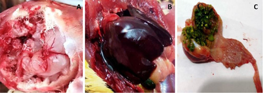
Post-mortem changes in naturally infected quails (2 months age) from Egypt: (A) Engorged blood vessels, congestion and hemorrhages in brain; (B) Severe congestion in the liver; (C) Greenish contents of the gizzard.

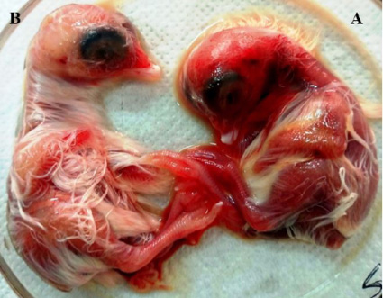
The inoculation of the supernatant of tissue suspension prepared from pooled organs of naturally infected quails into the allantoic cavities of 10-day-specific pathogen free-embryonated chicken eggs (A) Embryo showing congestion, dwarfism and sub-cutaneous hemorrhages on the head and legs and (B) The negative control embryo.
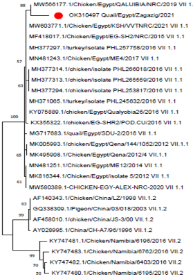
Phylogenetic analysis of NDV F gene fragment sequences through a bootstraps trail of 1000 was determined with the MEGA X using neighbor-joining method for tree construction. Our strain was labeled as red circle and was aligned with other strains of sub genotype.

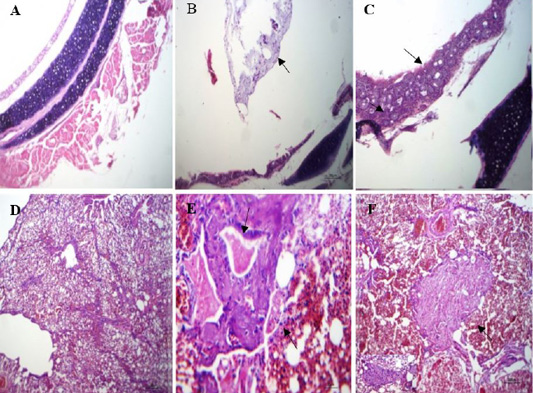
Histopathological lesions in the trachea and lungs of naturally infected quails: (A) trachea of normal quail (non-infected) showing normal wall and lumen, (HE, x100); (B) trachea of infected quail with NDV showing mucus exudate inside tracheal lumen(arrow), (HE, x100); (C) trachea of infected quail with NDV showing thickened tracheal wall by leukocyte(arrow) and proliferative glands (arrow head), (HE, x400); (D) lung of normal quail showing normal pulmonary tissue, (HE, x100); (E) lung of infected quail with NDV showing bronchial exudate (arrow), necrosed muscles (arrow head) and sero fibrinous fluid in adjacent air vesicle, (HE, x400);(F) lung of infected quail with NDV showing focal pneumonic area fibroblasts, macrophages and giant cells within pulmonary exudate (arrow), (HE, x100).

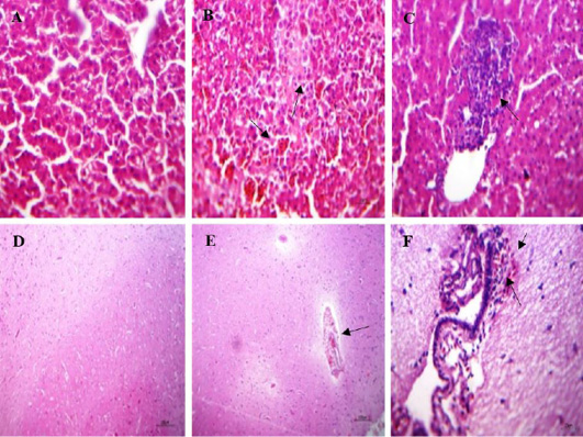
Histopathological lesions in the liver and brain of naturally infected quails; (A) liver of normal quail showing normal hepatic parenchyma, (HE, x400); (B) liver of infected quails with NDV showing intense sinusoidal congestion, large area of coagulative necrosis containing erythrocytes apoptosis(arrow) and minute necrosis of the hepatic parenchyma, (HE, x400); (C) liver of infected quails with NDV showing interstitial lymphocytic aggregation, (HE, x400); (D) brain of normal quail showing apparently normal brain tissue, (HE, x100) (E) brain of infected quails with NDV showing perivascular hemorrhages in Virchow robin spaces and non-suppurative encephalitis. besides, hemorrhages in meninges, (HE, x100); (F) brain of infected quails with NDV showing sub meningeal lymphocytic infiltration, extra vasated erythrocytes and edema, (HE, x400).

