
Seasonal Variation in the Microscopic Anatomy of Gonads and Gonadosomatic Index of Clupisoma garua


Seasonal Variation in the Microscopic Anatomy of Gonads and Gonadosomatic Index of Clupisoma garua
Riaz Hussain Pasha1*, Muhammad Zubair Anjum2, Imran Ullah2, Muhammad Akram Khan3, Adnan Ali4, Saif-Ur-Rehman5, Sana Batool2 and Arslan Emmanuel2

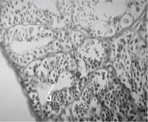
Figure 1
Preparatory Phase in the testis of Clupisoma garua during spring season. S: Spermatozoa.

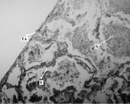
Figure 2
Spawning phase in the testis of C. garua during summer season. S: Spermatozoa, TA: Tunica albuginea.

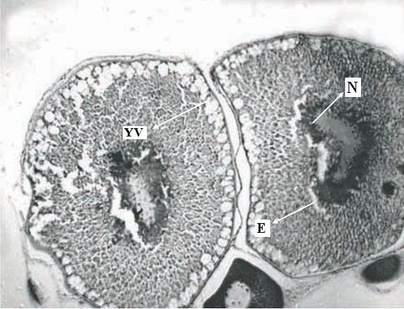
Figure 3
Preparatory phase in the ovary C. garua during spring season. N: Nucleus, E: Evagination in nucleus, YV: Yolk vesicles.
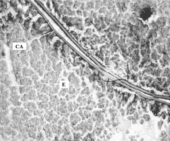
Figure 4
Spawning phase in the ovary of C. garua during summer season. E: Eggs, CA: Cortical Alveoli.
December 2019
Vol. 32, Iss. 4, Pages 562-709
