

Spermidine-Induced Autophagy Regulates the Survival of HeLa Cells


Spermidine-Induced Autophagy Regulates the Survival of HeLa Cells
Yihong Tian, Yongmei Qi*, Sajid Naeem, Ke Gao and Yingmei Zhang*
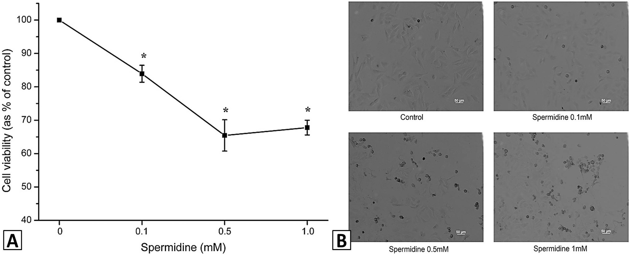
Spermdine reduced the viability of HeLa cells. Cells treated with spermidine (0, 0.1, 0.5, 1.0 mM) for 8 h. Cell viability was measured using the MTT assay. Results are expressed as the percentage of control and represent the mean ± S.E.M. of three independent experiments performed with triplicate cultures. *p < 0.05, compared with control (A). Images of cells were taken after exposure to spermidine as described above (B).
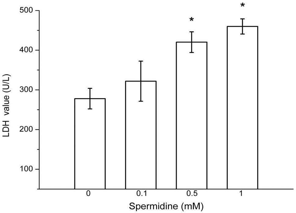
Spermdine increased the LDH leakage of HeLa cells. LDH levels in the supernatant was assessed using the LDH assay kit. Results are expressed as the percentage of control and represent the mean ± S.E.M. of three independent experiments. *p < 0.05, compared with control.

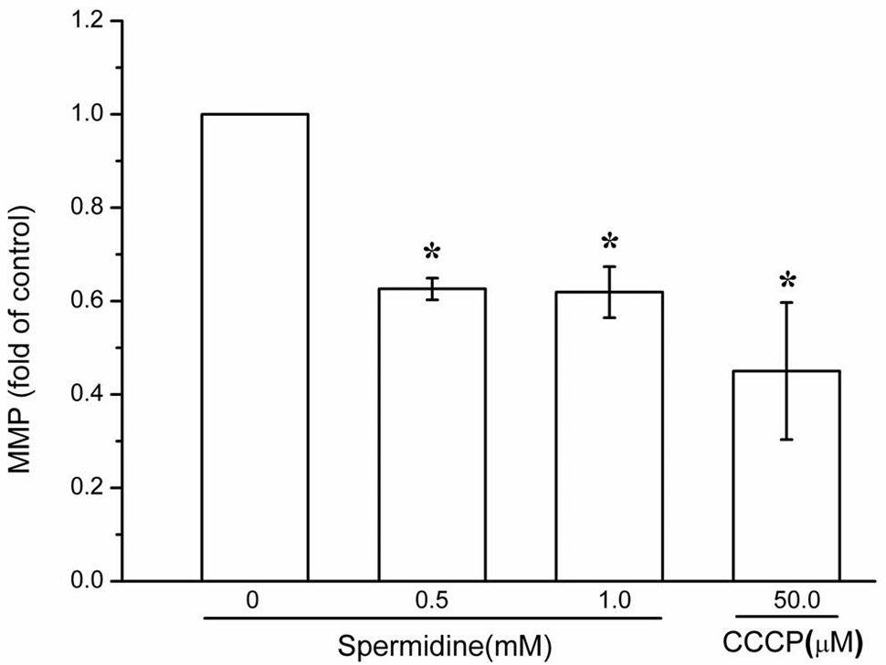
Spermdine collapsed MMP in HeLa cells. Cells were treated with spermidine (0, 0.5, 1.0 mM) for 8 h or 50 μM CCCP for 3 h. MMP was measured by flow cytometry. Results are expressed as the percentage of control and represent the mean ± S.E.M. of three independent experiments. *p < 0.05, compared with control.

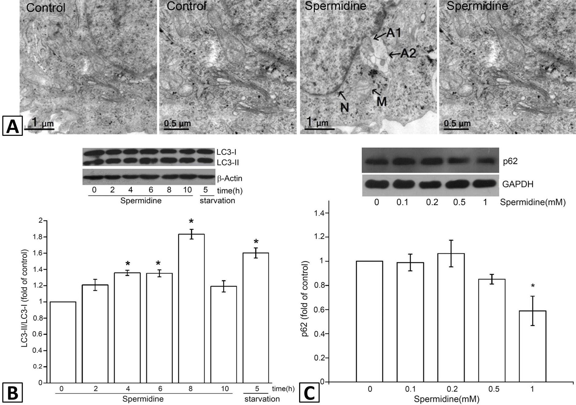
Spermidine induced autophagy in HeLa cells. HeLa cells were treated with 0.5 mM spermidine for 8 h. The formation of autophagic vacuole was observed under TEM (A). The nucleus is marked with letter N, letter M indicates intact mitochondria. autophagic vacuole is labeled as A1, A2. Cells were treated with 0.5 mM spermidine for 2, 4, 6, 8, 10 h or starved for 5 h. The expression of LC3 protein was assessed by western blotting and followed by grey scale analysis (B). Alternatively, the cells were treated with indicated concentrations of spermidine for 8 h and followed by western blotting of p62 (C). *p < 0.05, compared with control.

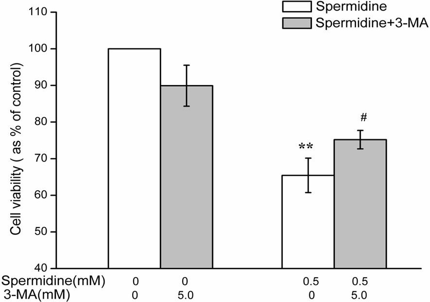
Spermidine-induced autophagy reduced viability of HeLa cells. Cells were treated with spermidine and 3-MA alone or simultaneously for 8 h. Cell viability was measured as in Figure 1. **p < 0.01, compared with control. #p < 0.05, compared with spermidine treatment alone.


