

L237P Substitution in the UROS Gene Causes X-linked Recessive Congenital Erythropoietic Porphyria in Pakistani Consanguineous Family Through Altered Uroporphyrinogen III Binding


L237P Substitution in the UROS Gene Causes X-linked Recessive Congenital Erythropoietic Porphyria in Pakistani Consanguineous Family Through Altered Uroporphyrinogen III Binding
Roshana Mukhtar1, Shaheen Shahzad1*, Sajid Rashid2, Maryam Rozi2, Madiha Rasheed3, Imran Afzal4 and Pakeeza Arzoo Shaiq5

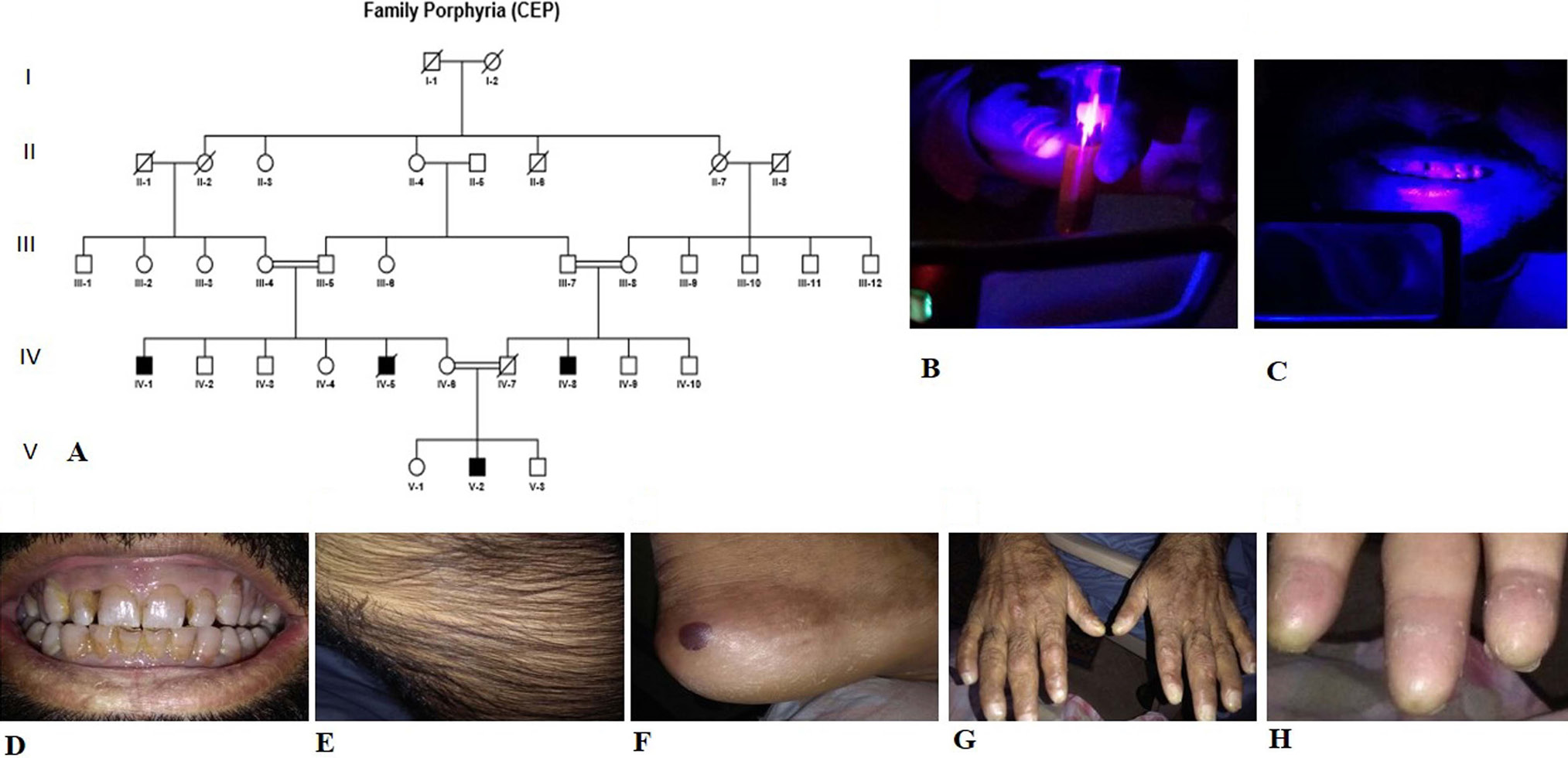
(A) Pedigree of the 5-generation consanguineous Pakistani family, showing features of X-linked recessive Congenital Erythropoietic Porphyria. Consanguineous unions are shown through double lines. Males and females are represented using squares and circles respectively. The affected and unaffected family members are shown using filled and clear symbols respectively, while a deceased individual is represented through a diagonal line. (B) Urine under wood lamp test. (C) Teeth colour under wood lamp test. (D-H) the clinical appearance of the affected persons (D) Eryhtrodondia, (E) Hypertrichosis, (F) Blisters formation on skin exposed to sunlight, (G) Blister healing with scarring, (H) Hands mutilating deformities of fingers with no finger prints.
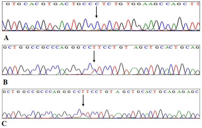
Sequence analysis of the UROS gene revealing the mutation c.935T>C (p.Leu237Pro). (A) shows the nucleotide sequence of an affected individual, (B) a heterozygous carrier, (C) a homozygous normal individual. The arrow represents the missense mutation.

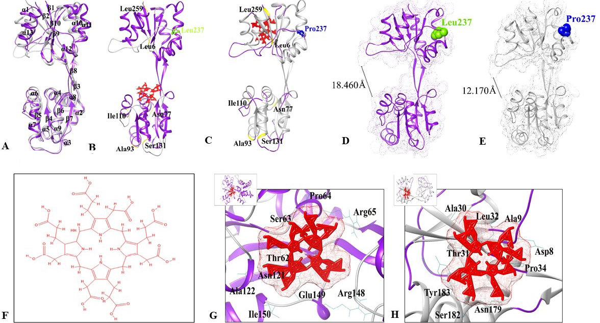
Structure analysis of UROSWT and UROSL237P. (A) Superimposition of UROSWT and UROSL237P structures. UROSWT is indicated in purple color, while UROSL237P is indicated in grey color. (B) Topview representation of UROSWT-Urogen complex in purple color. (C) UROSL237P -Urogen complex in grey color. Urogen is shown in red colored stick representation. Leu237 and Pro237 residues are indicated by green and blue spheres. (D-E) Structural comparison. (D) UROSWT is indicated in purple color, (E) UROSL237P model is indicated in grey color. Leu237 and Pro237 residues are indicated by green and blue spheres. Dotted lines indicate distance between adjacent residues. Bond length is indicated in Å. (F) Binding mode analysis of UROSWT and UROSL237P. (G) 2D structure of Urogen (3-[7, 12, 18-tris (2-carboxyethyl)-3, 8, 13, 17 tetrakis (carboxymeth-yl)-5, 10, 15, 20, 21, 22, 23, 24 octahydroporphyrin-2-yl] propanoicacid). (H) UROSWT (purple) binding with Urogen (red). (C) UROSL237P (grey) binding with Urogen (red). The interacting residues are labelled in black color with wire representation.

