

The Expression of IL-18Rα in Inferior Mesenteric Ganglion of Female Goat


The Expression of IL-18Rα in Inferior Mesenteric Ganglion of Female Goat
Yanjie Guo1, Weini Wu1, Hongmei Wang2, Xiao Guo2 and Yongping Xu2,*

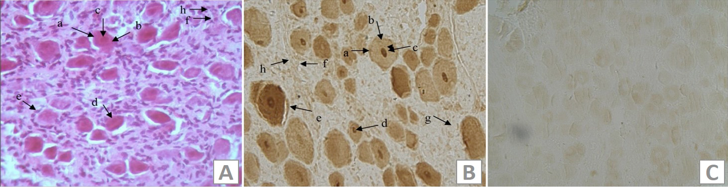
Immunohistochemical analysis of IL-18 Rα in IMG. A, the HE stains of the IMG of female goat; B, the immunohistochemical stains of IL-18Rα in the IMG; C, the blank control. a, membrane of IMG neuron; b, cytoplasm of the IMG neuron; c, nucleus of the IMC neuron; d, nucleoli of the IMC neuron; e, sertoli cells; f, blood vessels; g, schwann cell; h, endothelial cell.
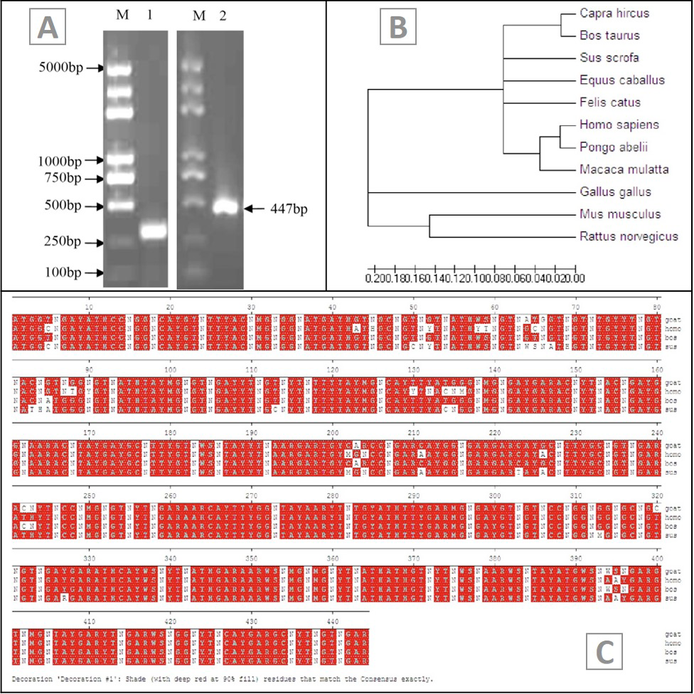
PCR analysis of IL-18Rα in IMG. A, agarose gel electrophoresis analysis of the PCR product of female goat IL-18Rα; M, DNA marker DL5000; Line 1, β-actin; Line 2, PCR product of IL18Rα; B, phylogenetic analysis of IL-18Rα; C, the alignment of goat IL-18 Rα between Homo sapiens, Sus scrofa and Bos taurus. Goat IL-18 Rα is 91%, 88% and 87% homologous to Bos taurus, Sus scrofa and Homo sapiens, respectively.
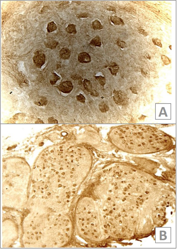
Analysis of IL-18Rα mRNA expression in IMG by in situ hybridization. Representative hybridization signals in IMG are brown (A, ×400; B, ×100).

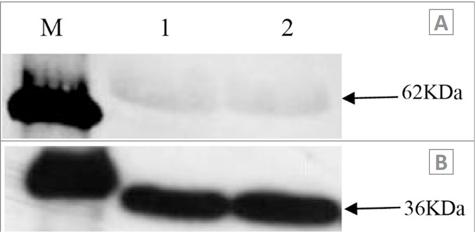
Analysis of IL-18Rα expression in IMG of female goat by Western blot. A, there is one band expected for gPR (about 62 kDa) was detected in the female goat IMG; B, the expression of β-actin; M, protein marker; 1 and 2, two repeats.

