

Construction and Characteristic Analysis of Omp10 Deletion Mutant of Brucella abortus


Construction and Characteristic Analysis of Omp10 Deletion Mutant of Brucella abortus
Tiansen Li1, Meiling Huang2, Zhen Wang1,Fei Guo3,Hui Zhang1,* and Chuangfu Chen1,*

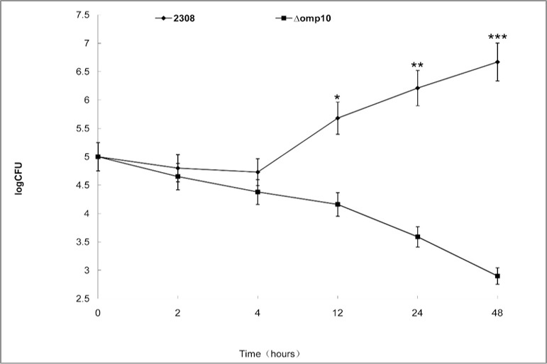
Intracellular survival of B. abortus 2308(♦), Δomp10 (■) mutants in murine RAW264.7 cells. Macrophages were infected with the strains, and incubation at 37 oC for the indicated time. Bacteria intracellular growth in RAW264.7 cells was determined at different incubation time points by counting the viable intracellular bacteria. Each point represents the mean±standard deviation of three experiments. Statistically significant differences between the bacterial growth of the parent strain and the mutants are indicated by asterisks (***, P<0.001).

Survival of B. abortus 2308 Δomp10 in the mouse model. Each group of mice was infected intraperitoneally with B. abortus 2308, Δomp10 mutant strains. Mice were infected by intraperitoneal injection with 1×10⁵ Brucella. The values are shown as means ± SEM of samples from 10 mice. Each point represents the mean±standard deviation of three experiments. Statistically significant differences between the bacterial growth of the parent strain and the mutants are indicated by asterisks (***, P<0.001).
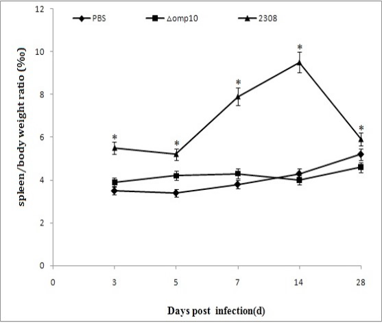
The changes of spleen weight/body weight at different time points post-infection. At different time points (3d, 5d, 7d, 14d, 28d) after infection, five mice were sacrificed, quickly removed the spleen and weighed. Statistically significant differences between the bacterial growth of the parent strain and the mutants are indicated by asterisks (***, P<0.001).

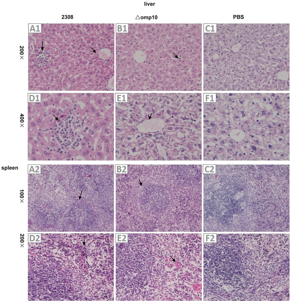
Virulence of B. abortus 2308 Δomp10 in the mouse model. Liver pathological section (A1-F1), the effect of 2308 on liver (A1, D1), the effect of Δomp10 on liver (B1, E1), the PBS control (C1, F1); Spleen pathological section (A2-F2), the effect of 2308 on spleen (A2,D2), the effect of Δomp10 on spleen (B2, E2), the PBS control (C2, F2).

Interaction of B. abortus 2308- and mutants-containing phagosomes with lysosomes in RAW264.7 macrophages. B. abortus were labeled with sheep anti-B. abortus IgG antibodies and Rhodamine (TRITC) conjugated AffiniPure donkey anti-sheep IgG antibody, lysosomes were labeled with Cell NavigatorTM Lysosomes Staining Kit Green Fluorescence. A, cells were fixed at different time points after infection. Confocal images of cells containing B. abortus were obtained at 4 h post-infection; B percentage of phagosome-lysosome fusion at different time points after infection. Fusion was evaluated by the colocalization of markers, Green fluorescein and TRD. To determine the percentage of fusion, bacteria were analyzed at each time point.
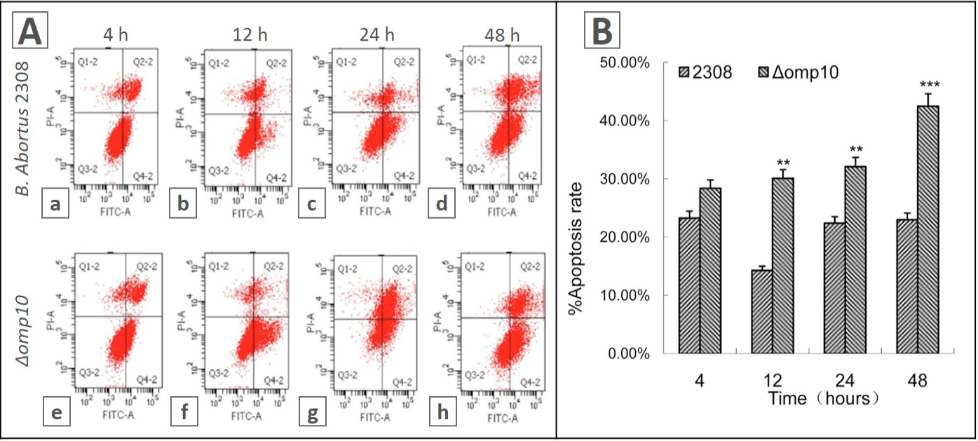
Flow cytometric analysis of RAW264.7 macrophages exposed to B. abortus 2308 and omp10 mutants. RAW264.7 macrophages were suspended with Binding Buffer and treated by Annexin V-FITC and PI at room temperature. A, cells were collected at different time points after infection, cell apoptosis images containing B. abortus. B, percentage of apoptosis at different time points after infection.


