

Distribution and Morphology of Ghrelin-Immunopositive Cells in the Testes of the African Ostrich


Distribution and Morphology of Ghrelin-Immunopositive Cells in the Testes of the African Ostrich
LX Ye1,2, JX Wang1,2,*, P Li1,2 and XT Zhang1,2
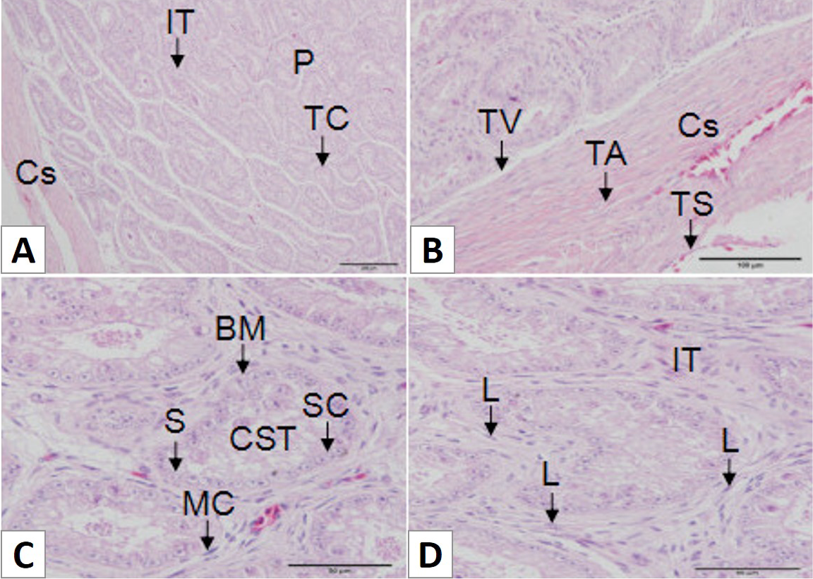
Histology of the African Ostrich testes. A, the testis was divided into the capsule (Cs) and parenchyma (P); B, the capsule was divided into three annular layers: tunica serosa (TS), tunica albuginea (TA) and tunica vasculosa (TV); C, the structure of the seminiferous tubules (CST) includes the basement membrane (BM), spermatogonia (S), Sertoli cells (SC) and myoid cells (MC); D, the structure of the interstitial tissue (IT) includes Leydig cells (L), tiny veins and connective tissue. Scale bar: 200 μm (A), scale bar: 100 μm (B) and scale bar: 50 μm (C and D).
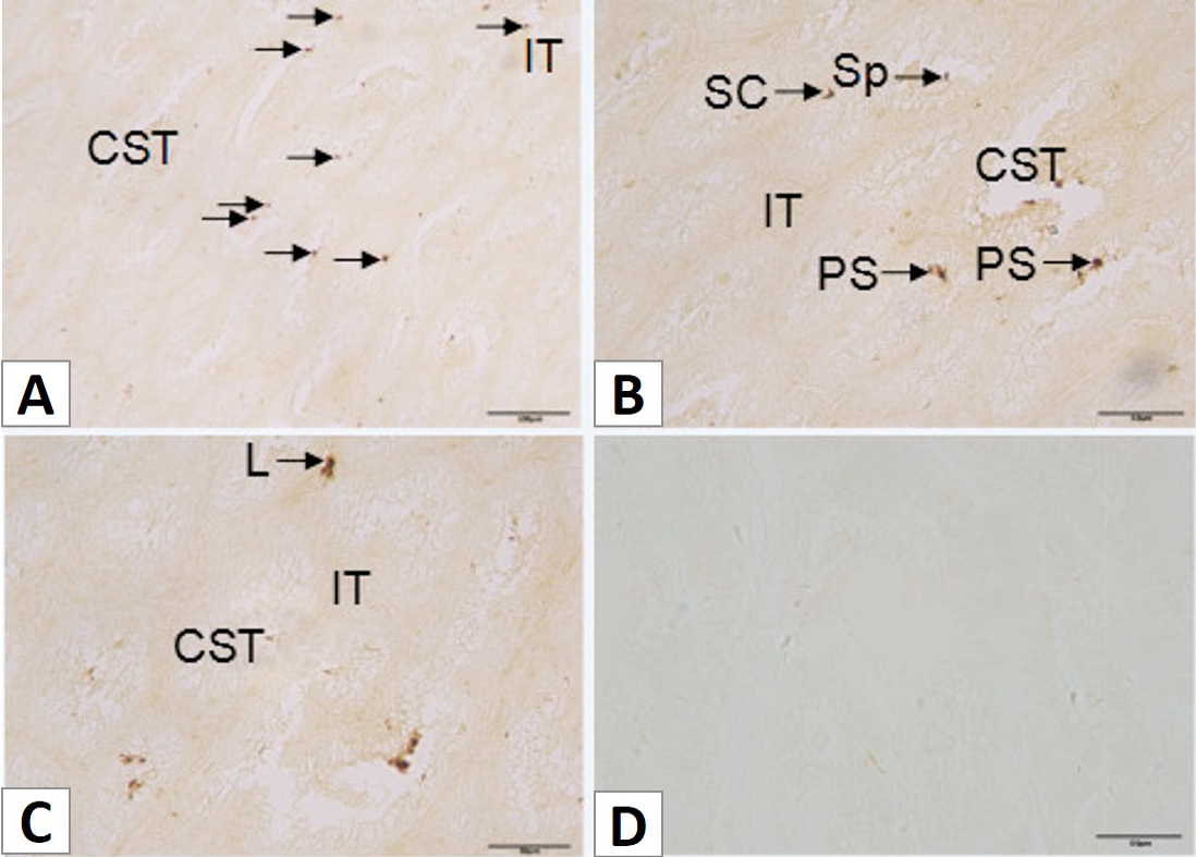
Distribution of ghrelin-ip cells in the testes of the African ostrich. A, ghrelin-ip cells were found in the seminiferous tubules (CST) of the tubular compartments and interstitial tissue (IT) of the testis; B, ghrelin-ip cells were found in the sperm (SP), primary spermatocytes (PS) and Sertoli cells (SC) of the seminiferous tubules; C, ghrelin-ip cells were found in the Leydig cells (L) of the interstitial tissue; D, microphotograph of absorption test in the testes. Arrows indicate ghrelin-ip cells. Scale bar: 100 μm (A) and scale bar: 50 μm (B, C and D).


