

Effect of Total Phenolic Acid on the Repeated Cerebral Ischemia-Reperfusion Model in Mice


Effect of Total Phenolic Acid on the Repeated Cerebral Ischemia-Reperfusion Model in Mice
Mingsan Miao*, Hui Zhao, Jiaojiao Jia, Xiaoyan Fang and Yanyan Miao
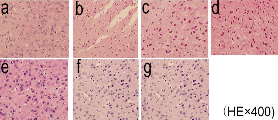
Effect of pathological changes in brain cortex of mice with repeated cerebral ischemia-reperfusion (HE×400). a. Blank Group; b. Model Group; c. Nimodipine Group; d. Naoluotong Group; e. High-dose Spatholobus Grandiflprum Group; f. Mid-dose Spatholobus Grandiflprum Group; g. Low-dose Spatholobus Grandiflprum Group.
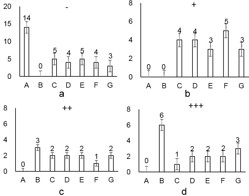
Effect of pathological changes in the cortex of brain tissue of mice with repeated cerebral ischemia-reperfusion. a: Cerebral cortical nerve cells are normal; b: Cerebral cortical individual nerve cell edema, individual neuron degeneration, cytoplasm light staining, fuzzy structure, individual neuron necrosis; c: Cerebral cortex a small number of nerve cell edema, scattered, a small number of neuronal degeneration The cytoplasm is lightly stained and the structure is fuzzy; d: Cerebral cortical nerve cells edema, most of the neurons are necrotic; A: Blank Group; B: Model Group; C: Nimodipine Group; D: Naoluotong Group; E: High-dose Spatholobus Grandiflprum Group; F: Mid-dose Spatholobus Grandiflprum Group; G: Low-dose Spatholobus Grandiflprum Group; -: Cerebral cortical nerve cells are normal; +: Cerebral cortical individual nerve cell edema, individual neuron degeneration, cytoplasm light staining, fuzzy structure, individual neuron necrosis; ++: Cerebral cortex a small number of nerve cell edema, scattered, a small number of neuronal degeneration The cytoplasm is lightly stained and the structure is fuzzy; +++: Cerebral cortical nerve cells edema, most of the neurons are necrotic.

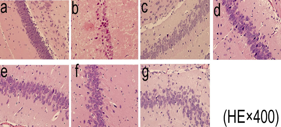
Effect of pathological changes in hippocampus of mice with repeated cerebral ischemia-reperfusion model (HE×400)

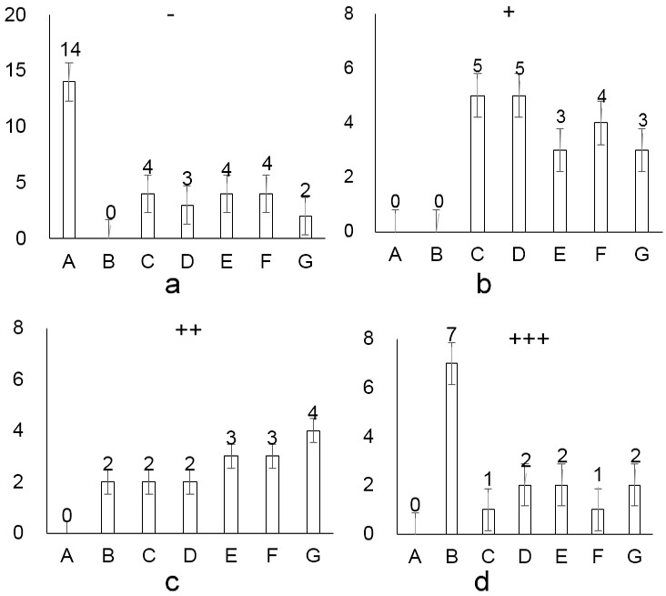
Effect of pathological changes in hippocampus of mice with repeated cerebral ischemia-reperfusion model; A: Blank Group; B: Model Group; C: Nimodipine Group; D: Naoluotong Group; E: High-dose Spatholobus Grandiflprum Group; F: Mid-dose Spatholobus Grandiflprum Group; G: Low-dose Spatholobus Grandiflprum Group; -: Brain cells in the hippocampus are normal; +: brain hippocampus, individual nerve cells edema, scattered, individual neuron degeneration, cytoplasm lightly stained, fuzzy structure; ++: a few hippocampal edema in the hippocampus of the brain, a few neuron degeneration, cytoplasm Light staining, fuzzy structure; ++: cerebral hippocampal neuronal edema, most of the neuron necrosis.


