

First Isolation of Brucella canis from Pet Dogs in Sichuan Province, China: Molecular Characterization, Pathogenicity and Antigen Location Analysis


First Isolation of Brucella canis from Pet Dogs in Sichuan Province, China: Molecular Characterization, Pathogenicity and Antigen Location Analysis
Zhijun Zhong1, Rui Tu1, Xichun Wang2, Yi Geng1, Qicheng Xiao1, Yinan Tian1, Bin Wei1, Jiaming Dan1, Ya Wang1 and Guangneng Peng1,*
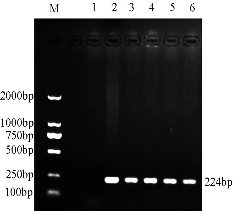
Brucella genus-specific PCR results of the strains isolated from five reference strains and two isolates from two pet dogs. Lane M, molecular weight marker; Lane 1, negative control with no DNA added; Lane 2, positive control with DNA extracted from the B. melitensis strain (16M); Lane 3, DNA extracted from isolate B. abortus (544A); Lane 4, DNA extracted from the B. suis (S2); Lane 5, DNA extracted from isolate strain W5; Lane 6, DNA extracted from isolate strain Y4.

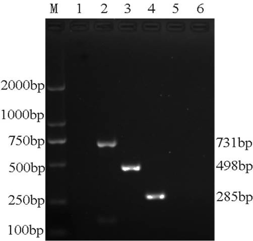
AMOS-PCR results of the strains isolated from three reference strains and two isolates from two pet dogs. Lane M, molecular weight marker; Lane 1, negative control with no DNA added; Lane 2, DNA extracted from B. melitensis (16M); Lane 3, DNA extracted from B. abortus strain (544A); Lane 4, DNA extracted from B. suis (S2); Lane 5, DNA of isolate strain W5; Lane 6, DNA of isolate strain Y4.

BcSS-PCR results of the strains isolated from three reference strains and two isolates from two pet dogs. Lane M, molecular weight marker; Lane 1, negative control with no DNA added; Lane 2, DNA extracted from the reference strain B. abortus (544A); Lane 3, DNA extracted from the reference strain B. melitensis (16M); Lane 4, DNA extracted from the reference strain B. suis (S2); Lane 5, DNA extracted from isolate strain W5; Lane 6, DNA extracted from isolate strain Y4.
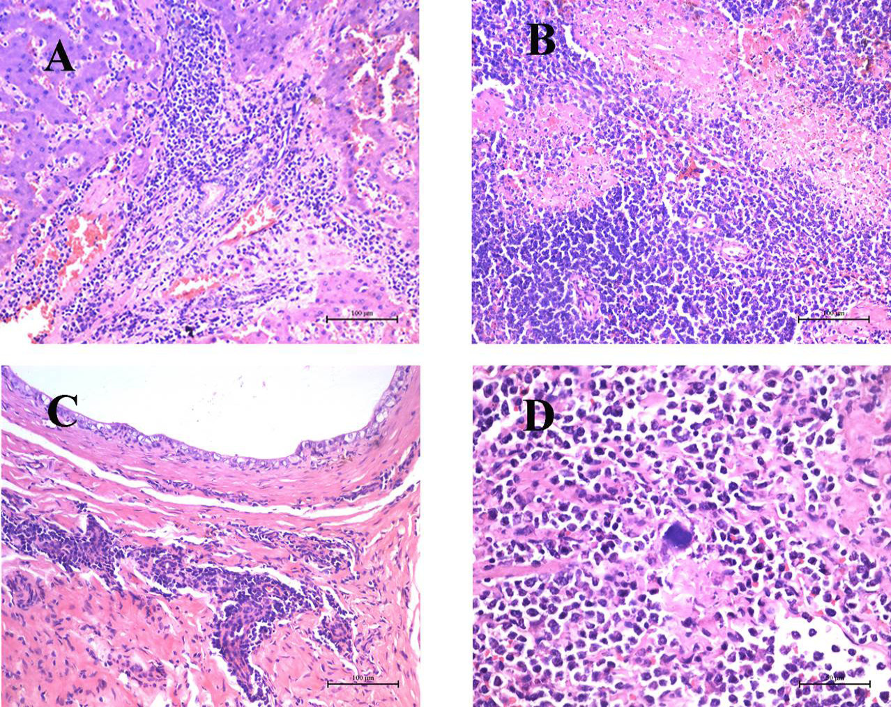
Pathologic lesions in liver, spleen, testicle and lymph nodes from the dog naturally infected with B. canis. A, liver showing severe infiltration of abundant lymphocytes and neutrophils in addition to hepatocellular necrosis and extensive vacuole degeneration of hepatocytes. HE Bar, 100 μm; B, spleen showing the presence of many granulomas with central necrotic areas in red pulp. HE Bar, 100 μm; C, testicle showing severe necrosis of spermatogenic cells and damaged seminiferous tubule structures accompanied by massive neutrophil, lymphocyte, and macrophage accumulation in interstitial tissue. HE Bar, 100 μm. D, lymph nodes showing proliferation of lymphocytes and reticuloendothelial cells along with deposition of fibrinous material; a bacterial bluish discoloration was found adjacent to the necrotic foci. HE Bar, 50 μm.
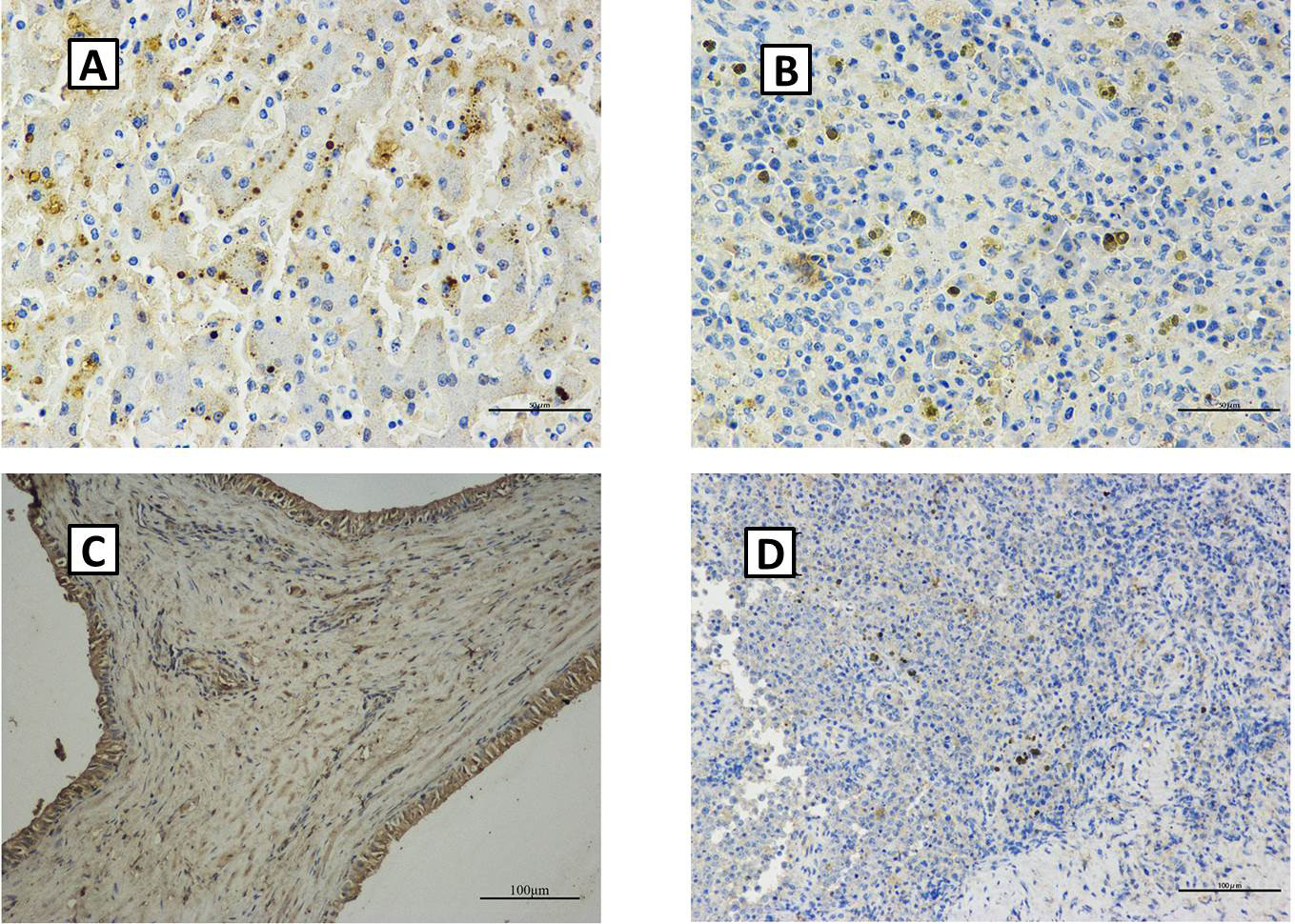
IHC staining of B. canis antigens in the lesions of four organs (liver, spleen, testicle, and lymph nodes). A, positive staining in the cytoplasm of macrophages, neutrophils and Kupffer cells of the liver (200×); B, Brucella antigens were detected in the cytoplasm of macrophages in the red splenic pulp (200×); C, B. canis antigens in the cytoplasm of interstitial macrophages and spermatogenic cells of the testicle (200×); D, positive staining in macrophages of the lymph nodes (200×).


