

Histological and Ultrastructural Features of the Leydig Cells and their Association with other Testicular Cells of the Vervet Monkey, Chlorocebus aethiops


Histological and Ultrastructural Features of the Leydig Cells and their Association with other Testicular Cells of the Vervet Monkey, Chlorocebus aethiops
Sogolo L. Lebelo1,* and Gerhard van der Horst2
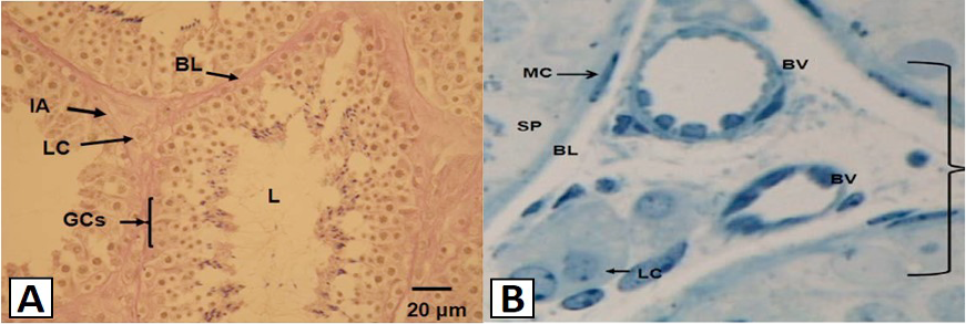
The light micrograph showing the general layout of the interstitial area (IA) and the seminiferous tubules (A), and Leydig cells (LC) and blood vessels (BV) of the testis (B) of the vervet monkey, Chlorocebus aethiops. A, Each tubule is surrounded by a basal lamina (BL). The Leydig cells (LC) are found in the interstitial area (IA). Within the seminiferous tubules are developing germ cells (GCs), which will later become the mature spermatozoa. The lumen (L) is occupied by spermatids of different stages; B, The myoid cells (MC) are found forming the basement membrane or the basal lamina (BL). On the basement membrane lies the spermatogonium (SP).
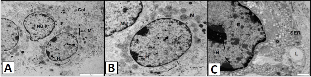
TEM micrograph showing Leydig cells of the vervet monkey, Chlorocebus aethiops. A, shows the polygonal nuclei (NU). The nucleus contains patches of euchromatin (E) and heterochromatin (H), with the latter situated at the periphery of the nucleus, close to the nuclear envelope. The cytoplasm contains numerous mitochondria (M) found closer the nucleus. The collagen fibers (COL) are also observed in the apical region of the cell. B, shows the round nuclei (NU) of the Leydig cells. The euchromatin (E) and heterochromatin (H) are observed in the nucleus. The cytoplasm contains numerous mitochondria (M) found closer the nucleus. C, shows the nucleus and the smooth endoplasmic reticulum (SER) of the Leydig cells. The nucleus contains euchromatin (E) and heterochromatin (H) patches. The lipid droplet (L) is found in association with the SER.
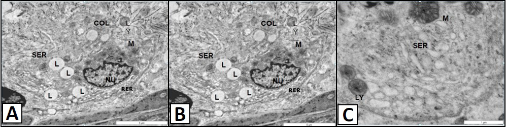
TEM micrograph showing of the Leydig cells of the vervet monkey, Chlorocebus aethiops. A, shows the polygonal nucleus (NU) has evenly distributed heterochromatin patches. The rough endoplasmic reticulum (RER) is situated at the basal region of the nucleus. The smooth endoplasmic reticulum (SER) is located at the apical region of the nucleus. The lipid droplet (L) and the lysosomes (LY) are found scattered in the cytoplasm together with collagen fibers (COL). B, show the distinct mitochondria of the Leydig cells. B, shows the smooth endoplasmic reticulum (SER) is located at the apical region of the nucleus. The lysosomes (LY) are found scattered in the cytoplasm. C, shows the tubular cristae (C) are clearly observed forming the mitochondria. The lipid droplets (L) are found in association with the mitochondria (M).
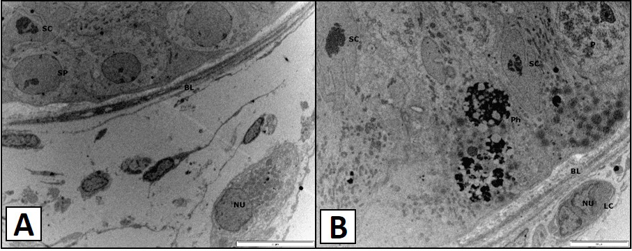
TEM micrograph showing the distinct nucleus (NU) of the Leydig cells of the vervet monkey, Chlorocebus aethiops. A, shows the Sertoli cell (SC) and the spermatogonium type B and found lying on the basal lamina (BL). B, shows the phagocytic vesicles are observed (Ph) and the spermatocyte at the pachytene (P) is observed. The Sertoli cells (SC) are found lying on the basal lamina (BL).


