

Ultrasonographic and Gross Pathological Studies on Testes and Epididymides of Rams and Bucks with Potential Lesions


Ultrasonographic and Gross Pathological Studies on Testes and Epididymides of Rams and Bucks with Potential Lesions
Saeed Murtaza1,*, Nazir Ahmad2, Muhammad Asif Raza3, Muhammad Saleem Akhtar1, Muhammad Mazhar Ayaz4, Muhammad Ali5 and Rais Ahmed6

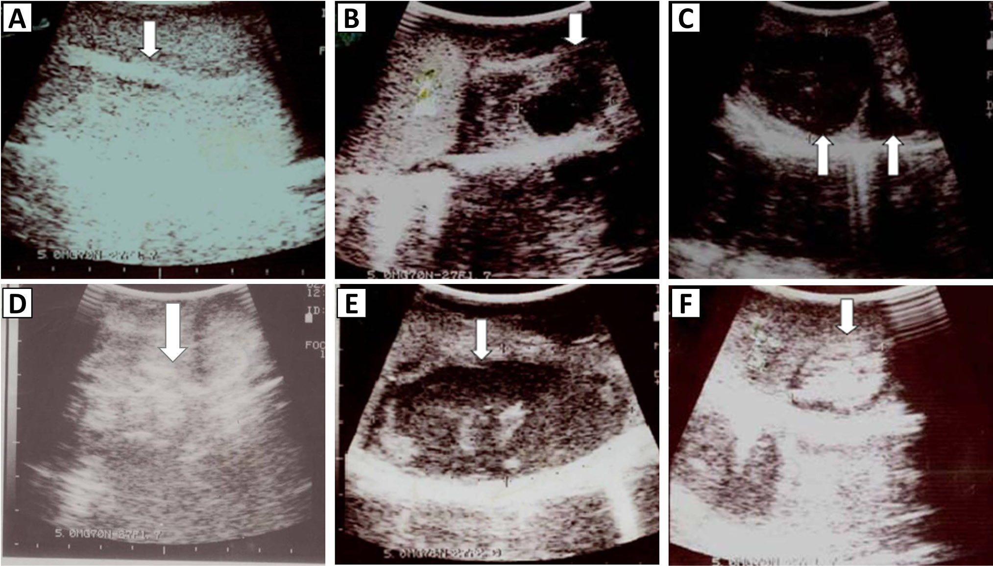
Longitudinal ultrasonograph of A, normal testis showing homogeneous and moderately echogenic parenchyma with hyperechoic mediastinum testis (arrow); B, showing sperm granuloma (arrow) at advance stage, having hyperechoic and anechoic areas; C, transverse ultrasonograph of the epididymal tail showing two sperm granulomata (arrows). Tissue below the lesion is hyperechoic, which is an ultrasound artifact known as enhanced through transmission; D, testis showing many hyperechoic areas scattered throughout the parenchyma (testicular mineralization); E, testicular abscess (arrow), showing hypoechoic and hyperechoic areas, with enhanced through transmission underneath the lesion (bright area); F, testis, showing a hyperechoic area surrounded by a hypoechoic line (arrow).


