

Honey and Black Seed Synergistically Promote Regeneration of Oligodendrocytes in Cuprizone Intoxicated Quail Brain


Honey and Black Seed Synergistically Promote Regeneration of Oligodendrocytes in Cuprizone Intoxicated Quail Brain
Mouhamed Zakiou Kolawole Adissa Raimi1, Ghulam Hussain2, Nousheen Zafeer3, Faheem Ahmad1, Muhammad Irfan4, Anwar Ullah1, Imdad Kaleem1 and Asghar Shabbir1*

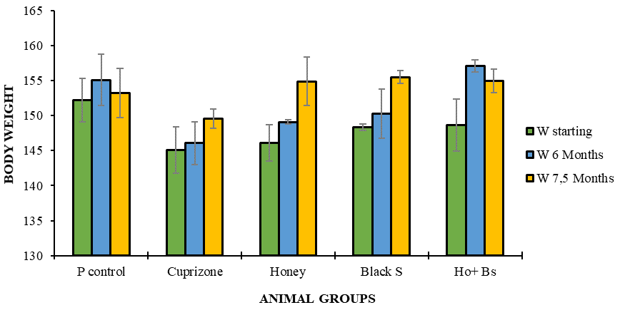
Body weights of animals at the starting, after six months and after seven and half months. There is no significant difference of body weight among the groups. Values are presented as mean t SEM. P value was set at 0.05.
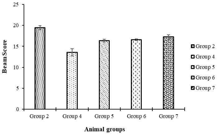
Beam walking score for five animal groups. Control group had significantly higher score as compared to all other groups. Honey, black seed and mixed groups showed significantly higher scores than CPZ treated animals. Scores are presented as mean ± SEM. Boxes with the same alphabet are not significantly different (P ≤ 0.05).

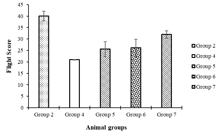
Flight length test results. Control group showed highest score followed by natural products treated groups and CPZ intoxicated group. Control group shows significantly higher length of flights than any other group. Treated groups have flight lengths less than control but significantly higher than animal treated with CPZ only. Data was presented as mean ± SEM. Bar with same alphabet means no significant difference (P ≤ 0.05).

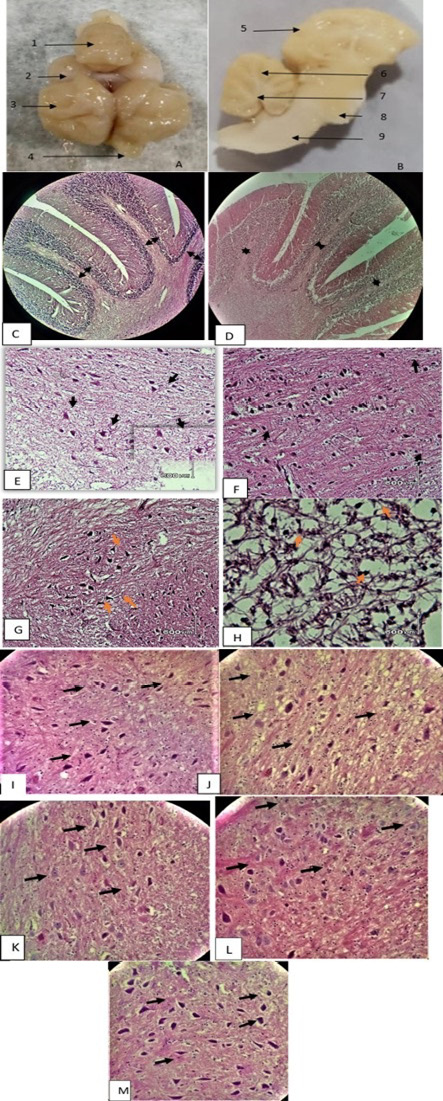
Cerebellar and cerebral white matter (H & E x 20 and x 40). (A) positive control entire brain (1: cerebellum; 2: optic lobe; 3: cerebral hemisphere; 4: olfactory lobe). (B) positive control sagittal brain section (5: cerebral cortex; 6: cerebellar cortex; 7: deep cerebellar white matter; 8: pons; 9: medullar oblongata. (C) positive control after six months shows normal thickness of cerebellar white matter folds. (D) six months CPZ treated animals show loss of cerebellar thickness in white matter folds. (E) control group kept for six months without CPZ shows deep cerebellar white matter and normal oligodendrocyte population. (F) animals with CPZ treatment for six months show rare oligodendrocytes. (G) cerebral white matter of control animal after six months shows normal glial cells. (H) cerebral white matter of six months CPZ treated animal shows apoptotic oligodendrocytes (arrow) and shrinkage of axons. (I) normal oligodendrocyte population can be seen in control animal after seven and half months without any treatment. (J) spontaneous oligodendrocyte regeneration can be seen in animal with no treatment for six weeks after six months of CPZ treatment. (K) honey treated group showing regeneration of oligodendrocytes. (L) black seed treated group showing increase in glial cells population. (M) Mixture (honey and black seed) treated group shows significantly increased oligodendrocyte population.
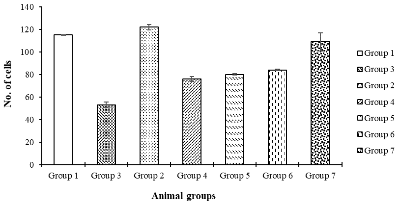
The number of oligodendrocytes in cerebellar white matter of quail is depicted in the graph. There is a severe depletion of oligodendrocytes after six months of CPZ treatment. Oligodendrocyte no. increases after the arrest of treatment. A non-significant difference in the number of oligodendrocytes can be observed in CPZ, honey and black seed group. Animals in mixture group have significantly higher number of oligodendrocytes than the honey, black seed and CPZ group. Oligodendrocyte number has been presented as mean ± SEM. Bar with same alphabet means no significant difference (P ≤ 0.05).

