

In Vivo Evaluation of purD Gene Deleted Brucella abortus in Mice as Potential Vaccine Candidate for Control of Brucellosis


In Vivo Evaluation of purD Gene Deleted Brucella abortus in Mice as Potential Vaccine Candidate for Control of Brucellosis
Muhammad Ilyas Riaz1, Masood Rabbani1*, Sohail Raza1, Ali Raza Awan2, Aleena Kokab1, and Rida Haroon Durrani1

Construction and confirmation of purD gene deletion plasmid cassette “pUC19-Kanamycin-Up-Down”. (A) Illustration of map of purD gene deletion plasmid cassette pUC19-Kanamycin-Up-Down (A1) confirmation of pUC19-Kanamycin-up-down gene deletion plasmid cassette (B) illustration of map of pUC19 plasmid (B1) confirmation of pUC19 cloning in DH5α cells (C) illustration of Kanamycin fragment with restriction sites KpnI (GGTACC) and BamHI (GGATCC) (C1) amplification of Kanamycin fragment. (C2) Confirmation of Kanamycin cloning in pUC19 (D) Illustration of Up sequence fragment with restriction sites EcoRI (GAATTC) and KpnI (GGTACC) (D1) amplification of upstream fragment (D2) confirmation of up sequence cloning into pUC19-Kanamycin plasmid (E) Illustration of downstream fragment with restriction sites BamHI (GGATCC) and PstI (CTGCAG) (E1) amplification of downstream fragment (E2) confirmation of Downstream fragment into pUC19-Kanamycin-up.
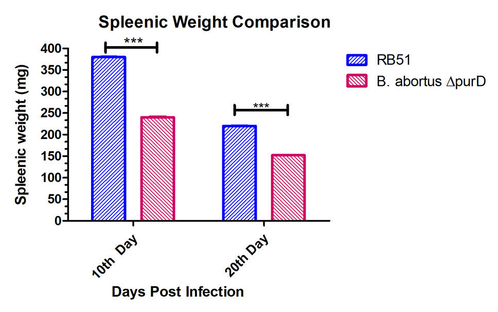
Comparison of splenic weight between RB51 and B. abortus ΔpurD mutant mice groups. Splenic weight comparison of the RB51 and B. abortus ΔpurD mutant mice groups infected with 3 X10⁸ CFU/ dose of the RB51 and B. abortus ΔpurD mutant at 10th and 20th DPI. Error bars represent average values ± the standard deviations. The asterisks denote values that are significantly different (*P <0.05) between groups with the parent RB51 and the B. abortus ΔpurD mutant at each time point, as determined by two away ANOVA.
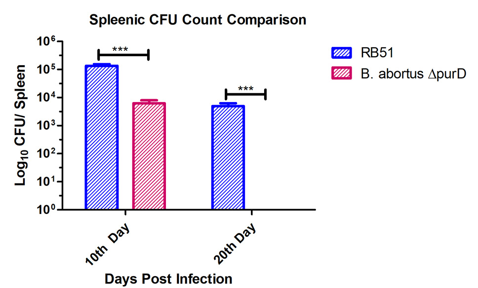
Comparison of Splenic CFUs between RB51 and B. abortus ΔpurD mutant mice groups. Clearance of the RB51 and B. abortus ΔpurD mutant in mice. Recovery of viable bacterial CFUs count from spleens of mice at 10th and 20th DPI. Results are expressed as total CFU per spleen. Error bars represent average values ± the standard deviations. The asterisks denote values that are significantly different (*P <0.05) between groups with the parent RB51 and the B. abortus ΔpurD mutant at each time point, as determined by two way ANOVA.

Representative images of histopathology of the RB51 and B. abortus ΔpurD mutant in mice. Six out of eight mice from RB51 mice group spleen showed atrophy of follicular area and dead tissue mass is also seen in the (arrow) with few giant cells are also present (circle) (A). Six mice out of eight from B. abortus ΔpurD mice group spleen showed giant phagocytic cells with phagocytized bacteria were seen (circle) and higher infiltration of lymphocytes (arrow) (B). Control mice group spleen showed no tissue changes and almost normal tissue parenchyma was seen (C).

