

Modulation of Tau Expression in the Colonic Muscle Layer of a Rat Model with Opioid-Induced Constipation


Modulation of Tau Expression in the Colonic Muscle Layer of a Rat Model with Opioid-Induced Constipation
Jinzhao Li1, Yawen Zhang2, Binghan Jia1, Yuqiong Zhao1, Huijuan Luo1, Xiaojie Ren1, Yuan Li3, Xiaoyan Bai1, Jing Ye3 and Junping Li1*

Body weight, fecal grade scores and intestinal propulsion velocities of rats in the 3 groups were estimated. A, Changes in body weight of rats in the 3 groups. B, Fecal traits of rats in the 3 groups. C, Fecal trait grade scores of rats in the 3 groups. D-F, Comparison of intestine propulsion velocity.

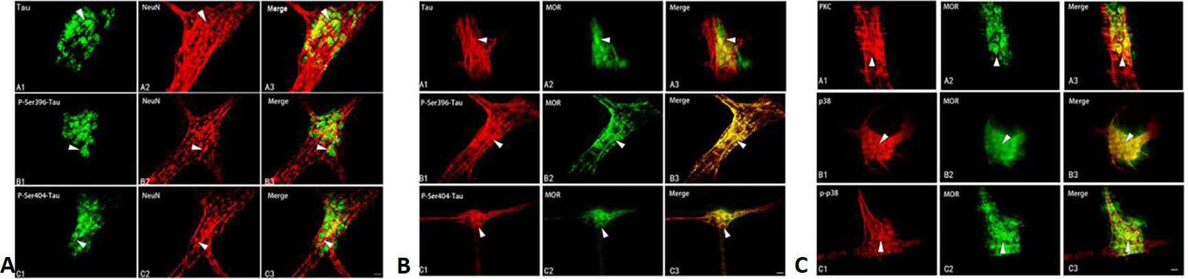
Immunohistochemistry of the colonic myenteric plexus tissues from normal rats. A, (A1) Tau-positive cells; (A2) NeuN-positive cells; (A3) Intracellular co-expressions of Tau and NeuN. (B1) P-Ser396-Tau-positive cells; (B2) NeuN-positive cells; (B3) Intracellular co-expressions of P-Ser396-Tau and NeuN. (C1) P-Ser404-Tau-positive cells; (C2) NeuN-positive cells; (C3) Intracellular co-expressions of P-Ser404-Tau and NeuN. B, (A1) Tau-positive cells; (A2) MOR-positive cells; (A3) Co-expressions of Tau and MOR markers in enteric neural cells. (B1) P-Ser396-Tau-positive cells; (B2) MOR-positive cells; (B3) Co-expressions of P-Ser396-Tau and MOR in enteric nerve cells. (C1) P-Ser404-Tau-positive cells; (C2) MOR-positive cells; (C3) P-Ser404-Tau and MOR double-positive enteric nerve cells. C, (A1) PKC-positive cells; (A2) MOR-positive cells; (A3) Co-expressions of PKC and MOR in enteric nerve cells. (B1) P38-positive cells; (B2) MOR-positive cells; (B3) Co-expressions of p38 and MOR in enteric nerve cells. (C1) P-p38-positive cells; (C2) MOR-positive cells; (C3) Co-expressions of p-p38 and MOR in enteric nerve cells. Scale bars: 20µm.


ELISA of protein content and statistical analysis of colonic muscular layer in the three groups of rats 7 days after modeling. A and B, comparisons of tau expressions at each phosphorylation site in the distal colonic muscular layer of NCG and OIC rats. C-F, the expressions of P-Ser396-Tau, P-Ser404-Tau, MOR and p-p38 were significantly increased in the colonic muscular layer of OIC rats compared with that of NCG and NSG (***P < 0.001).

Expressions of Tau, P-Ser396-Tau P-Ser404-Tau, MOR, PKC, p38 and p-p38 in the distal colonic muscles of three groups of rats on the 7th day of modeling. A-C, Protein expressions of Tau, P-Ser396-Tau, P-Ser404-Tau, MOR, PKC, p38 and p-p38. D-J, Statistical analysis of Tau, P-Ser396-Tau, P-Ser404-Tau, MOR, PKC, p38 and p-p38 expressions.
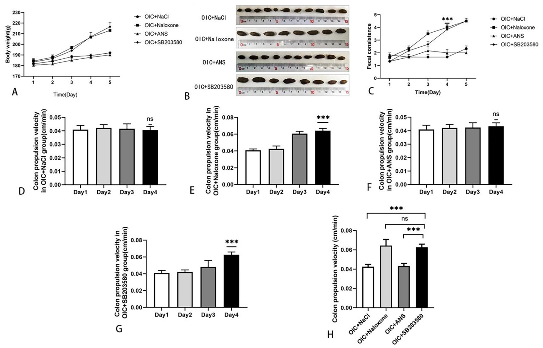
Comparisons of body weights, fecal trait grade scores of rats and colonic propulsion velocities in the four different groups following the drug intervention in vivo. A, Changes in the body weight of rats during drug intervention. B, Fecal traits of rats on the 4th day of drug intervention. C, Comparison of fecal trait grade scores during the drug intervention. D-G, Comparison of colonic propulsion velocity in four groups during 4 days of drug intervention. Compared with the 3rd day before drug intervention. H, Comparison of colonic propulsion velocities between the four groups of rats on the 4th day after in vivo drug intervention.

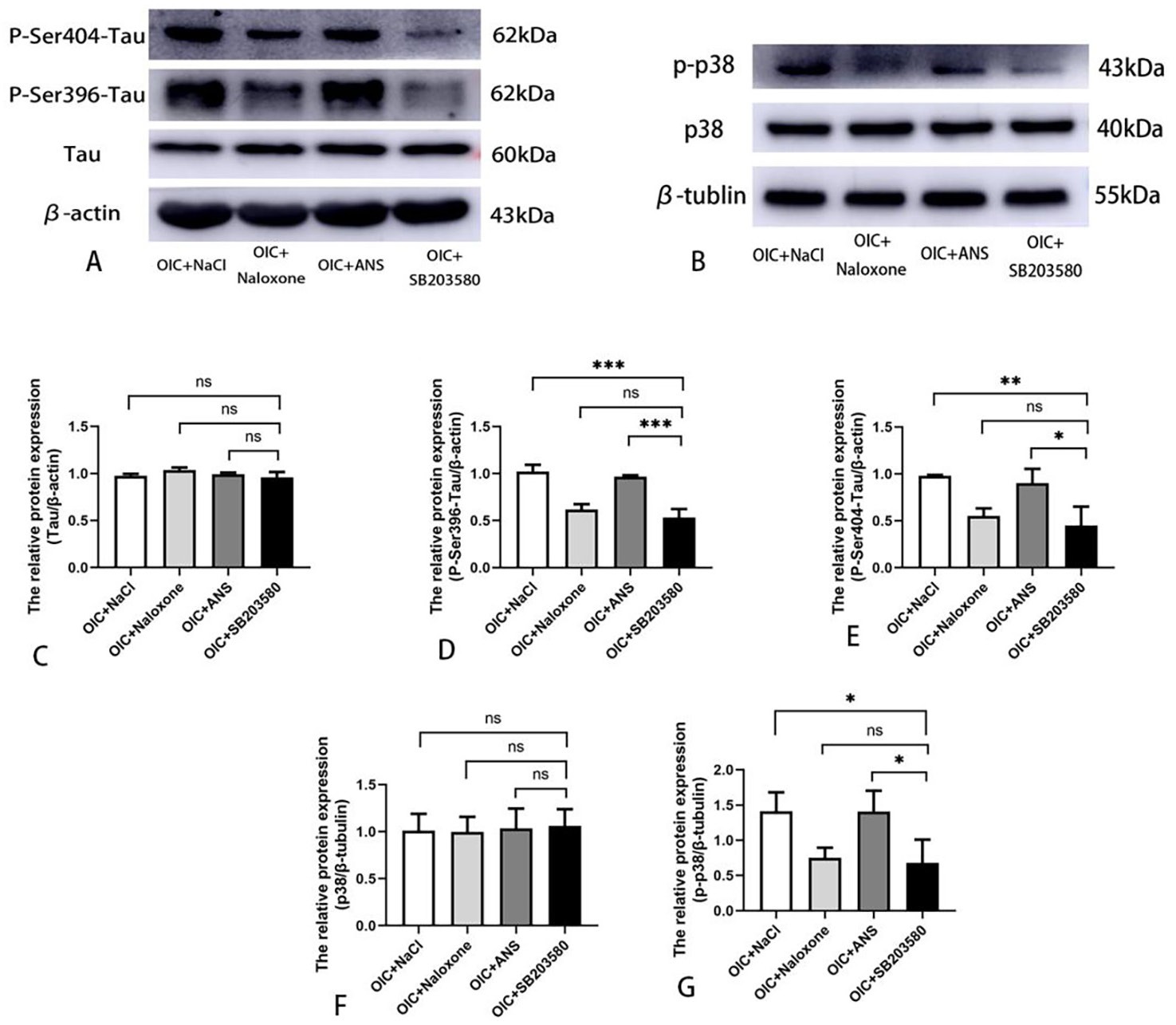
Changes in the expressions of Tau, P-Ser396-Tau, P-Ser404-Tau, p38 and p-p38 proteins in the distal colonic musculatures of four groups of rats after 4 days of in vivo drug intervention. A and B, Protein expressions of Tau, P-Ser396-Tau, P-Ser404-Tau, p38 and p-p38. C-G, Statistical analysis of Tau, P-Ser396-Tau, P-Ser404-Tau, p38 and p-p38 expressions.

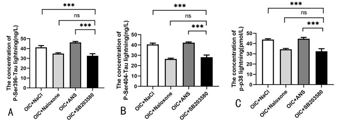
ELISA detection of protein contents and statistical analysis of colonic muscular layer in the four groups of rats after 4 days of intervention in vivo. A-C, On the 4th day of the imposed intervention, expressions of P-Ser396-Tau, P-Ser404-Tau and p-p38 were significantly reduced in the colonic muscular layers of rats in the OIC+SB203580 group compared with those in the OIC+NaCl and OIC+ANS groups (***P<0.001).

