

Alantolactone Suppresses YAP Signaling and Stemness Properties in Glioblastoma Cells


Alantolactone Suppresses YAP Signaling and Stemness Properties in Glioblastoma Cells
Bing Yan1, Meng Meng Zhang2, Xi Chen2, Yongming Li2*, Muhammad Khan3* and Tonghui Ma1*
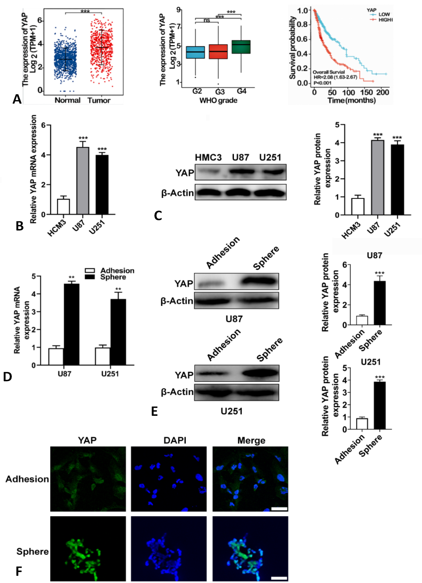
YAP is highly expressed in glioma cells and glioma stem cells. (A) Higher expression of YAP in gliomas (n =689) than in normal brain tissue (n = 1157) was obtained from the TCGA database (***P < 0.0001). Median and interquartile ranges are indicated by black lines. According to WHO grading criteria, (n=635, G2=224, G3=243, G4=168). Medians with interquartile spacing are indicated by black lines. Gliomas (n =697patients) were evaluated with YAP mRNA levels and the results were correlated with an overall survival of 18 years. The red line indicates patients with high YAP transcripts (n = 349) and the blue line indicates patients with low YAP transcripts (n = 348). P values were analyzed by Kaplan-Meier analysis using the GraphPad prism. (B) YAP expression levels were detected in Human Microglia Clone (HCM3) and human glioma cancer cells (U87 and U251) by qRT-PCR. *P < 0.05, **P < 0.01. (C) YAP expression levels were detected in Human Microglia Clone (HCM3) and human glioma cancer cells (U87 and U251) by Western blotting *P < 0.05, **P < 0.01. (D) mRNA expression levels of YAP in spheroid cells and adherent cells. (E) Protein expression of YAP in spheroid cells and adherent cells. (F) Immunofluorescence staining of YAP in spheroid cells and adherent cells in U87 cells (scale bar = 50μm).
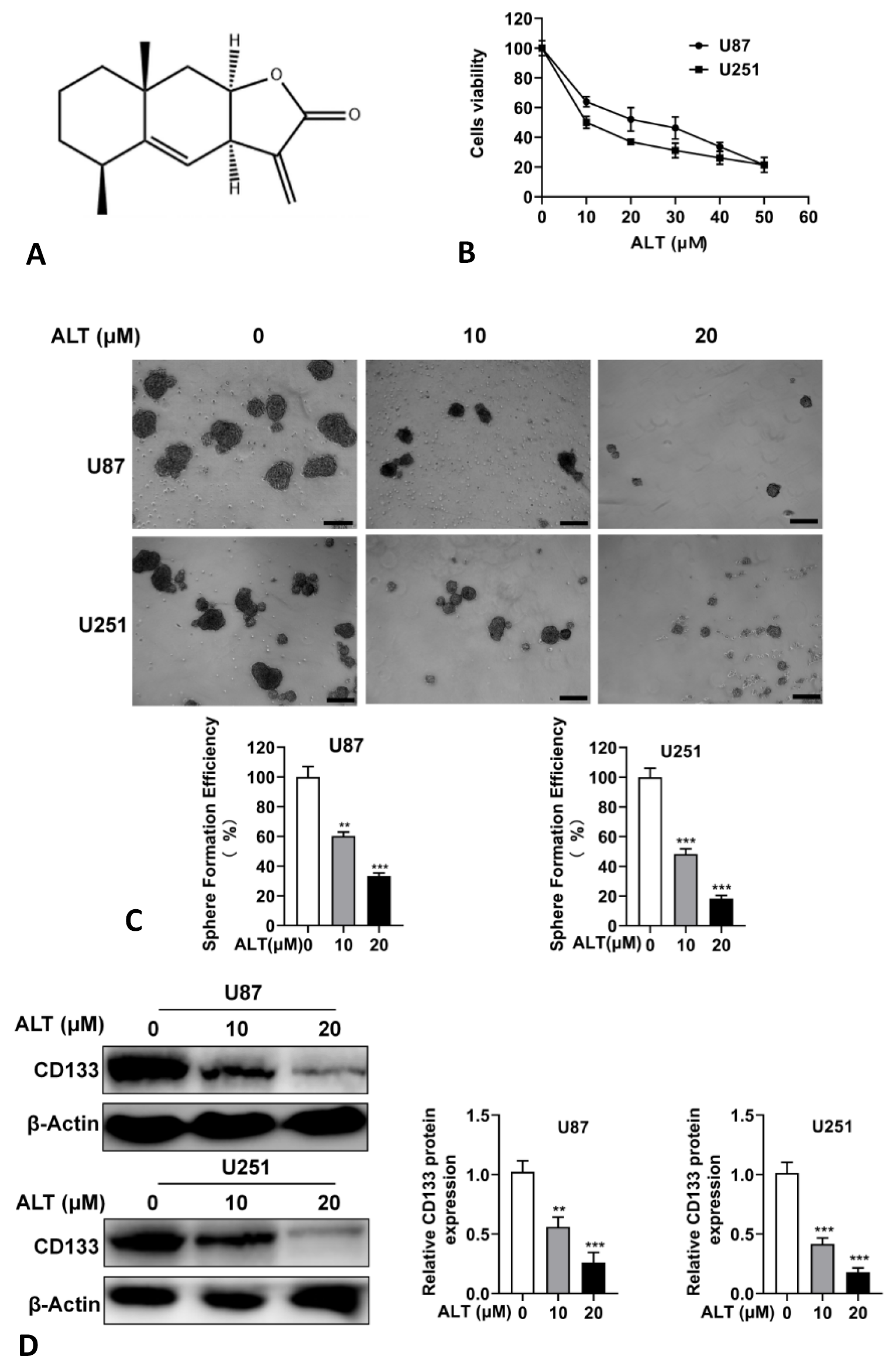
ALT suppresses stemness properties of glioma. (A) Chemical structure of ALT. (B) U87 and U251 cells were treated with ALT for the 48 h and CCK8 assay was performed. (C) The cells were treated with ALT for 96 h and mammosphere formation was measured. Pictures were taken under a reverse microscope (scale bar = 200 μm). (D) U87- and U251-spheroid cells were treated with ALT for 96 h, and protein expressions level of glioma stem cells marker gene CD133 was analyzed by Western blotting.

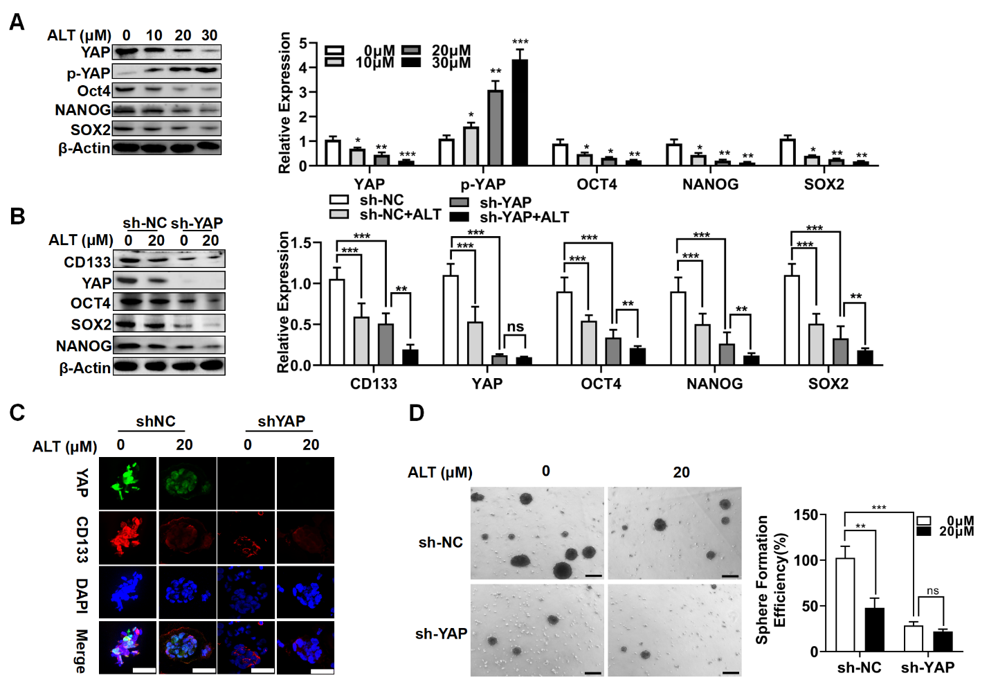
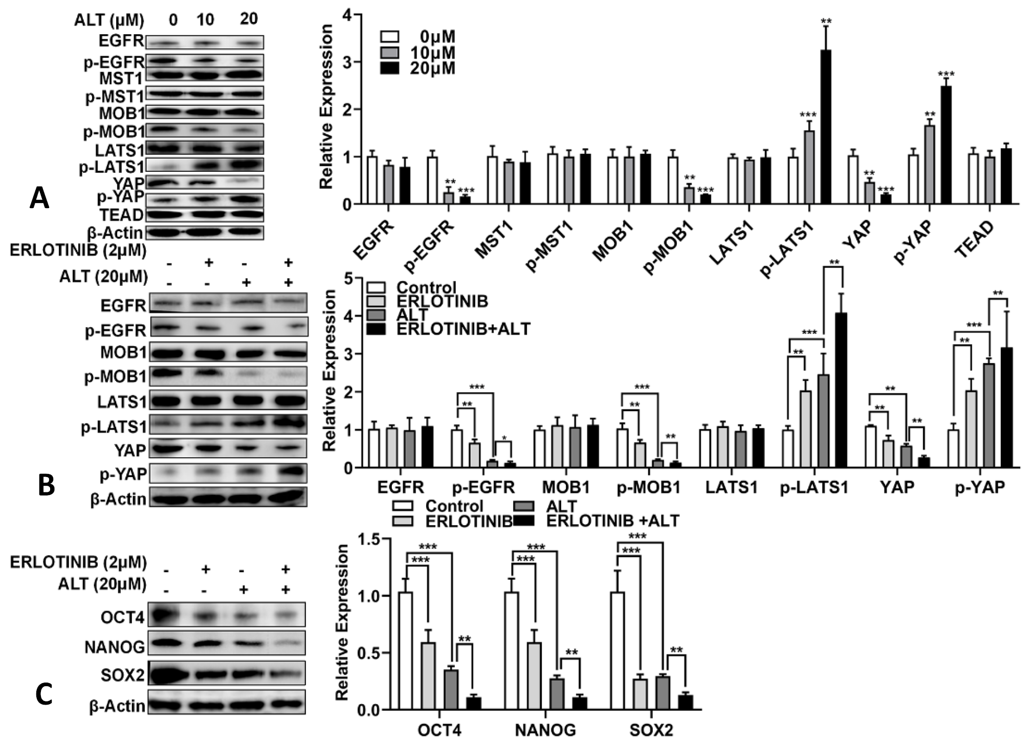
ALT suppresses stem-cell-like properties of glioma CSCs via suppressing YAP expression. (A) Western blot was used to examine the expression of stem cell markers (SOX2,OCT4,NANOG) and YAP, p-YAP in U87-spheroid cells treated with ALT for 96 h. (B) U87spheroid- shScramble cells and U87spheroid - shYAP cells were treated with ALT for 96 h, and expression of CD133,YAP, OCT4, SOX2 and NANOG was measured by Western blotting. (C) U87spheroid-shScramble cells and U87 spheroid - shYAP cells were treated with ALT (20μM) for 96 h, and expression of CD133 and YAP was observed by immunofluorescence (scale bar = 50 μm). (D) Comparison of mammosphere formation between U87-shScramble or U87-shYAP cells treated with ALT for 96h. (Scale bar =200 μm).
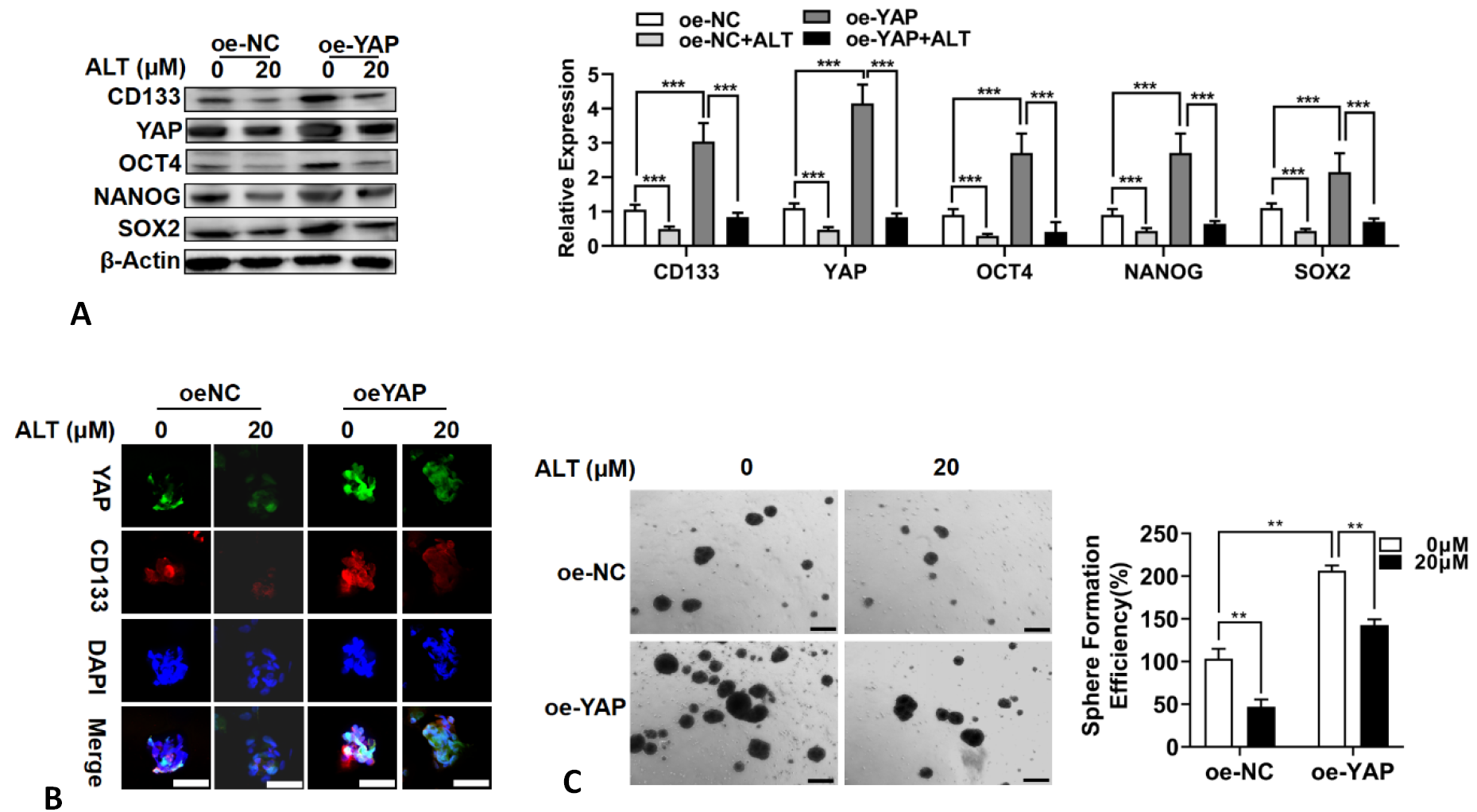
ALT suppresses stem-cell-like properties of glioma stem cells via suppressing YAP expression. (A) U87 spheroid- oeScramble cells and U87 spheroid- oeYAP cells were treated with ALT for 96 h and expression of CD133, YAP, OCT4, SOX2 and NANOG was measured by Western blotting. (B) Immunofluorescence was used to detect the expression of CD133 and YAP in U87spheroid-oeScramble cells and U87spheroid -oeYAP cells (scale bar =50 μm). (C) Comparison of mammosphere formation between U87-oeScramble or U87-oeYAP cells were treated with ALT for 96 h. (Scale bar = 200 μm).

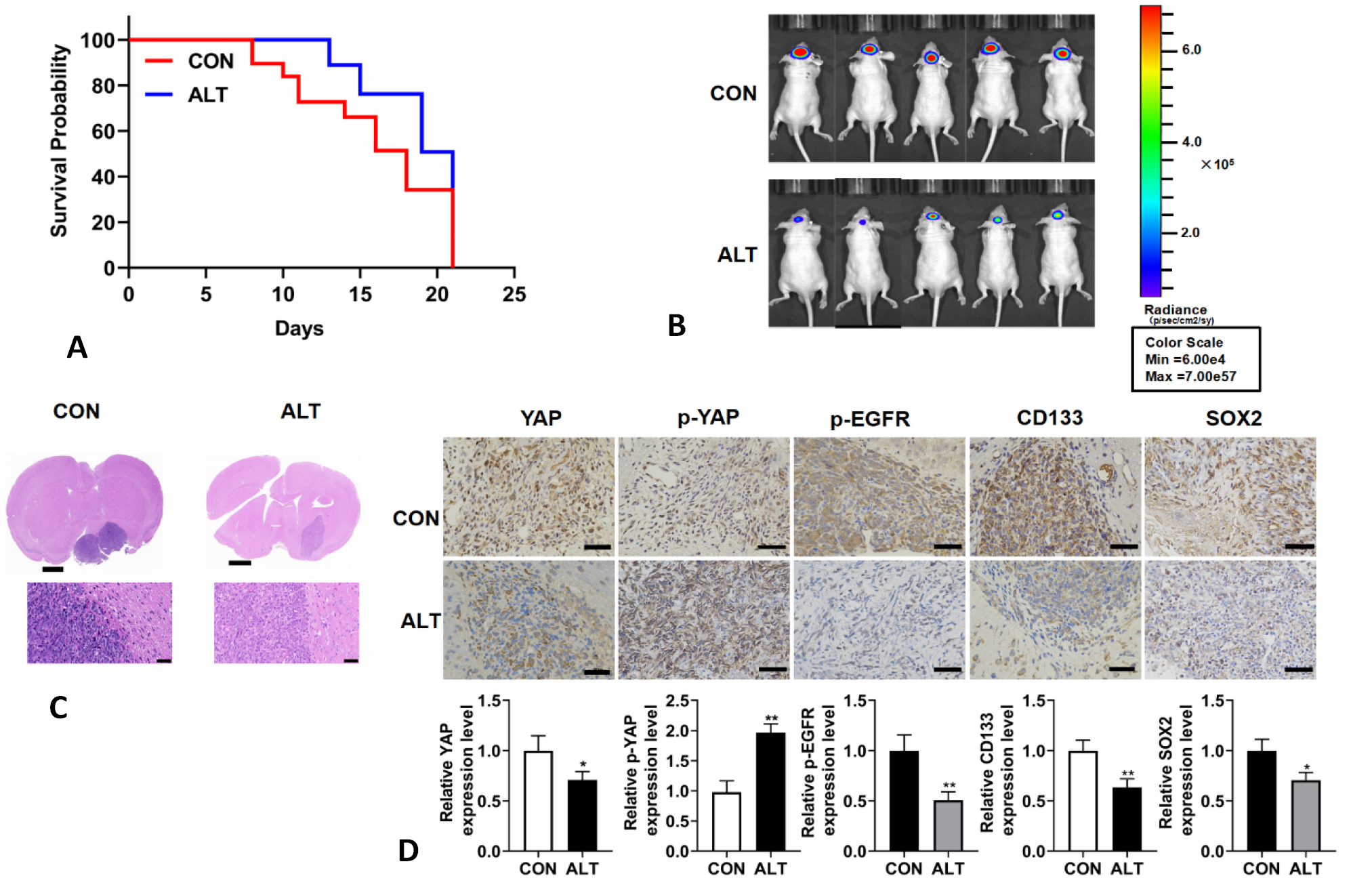
ALT inhibits the growth of glioma xenografts. (A) Survival analysis for animals treated with 0.9% saline or ALT (20mg/kg) (P< 0.01 by log-rank test; n 0.9% saline =12, n ALT =12). (B) Bioluminescence imaging showed the tumor size. (C) H and E staining of sections from mouse brains with U87 xenografts treated with 0.9% saline or ALT (20mg/kg) at 21 days. (D) IHC for YAP, p-YAP, p-EGFR, CD133 and SOX2 in sections from indicated xenografts (scale bar = 100 μm).

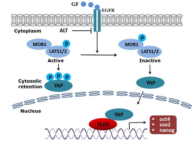
A schematic diagram representing intervention of Alantolactone with EGFR-mediated YAP signaling pathway and stemness properties. In the absence of EGFR ligand (EGF), LATS1/2 is phosphorylated (active). Upon activation, LATS1/2 induces YAP phosphorylation which ultimately results in cytoplasmic retention or degradation of YAP. Upon ligation with growth factor, EGFR become activated leading to MOB1 tyrosine phosphorylation and LATS1/2 dephosphorylation (inactive). Inactivation of LATS1/2 results in translocation of YAP into nucleus where it interacts with TEAD transcriptional factor and induces the expression of various genes associated with stemness properties of cell.


