
An Update in Histopathology and Immunohistochemistry of Ovarian Sex Cord-Stromal Camel Tumors


An Update in Histopathology and Immunohistochemistry of Ovarian Sex Cord-Stromal Camel Tumors
Ibrahim Elmaghraby*, Abdel-Baset I. El-Mashad, Shawky A. Moustafa, Aziza A. Amin

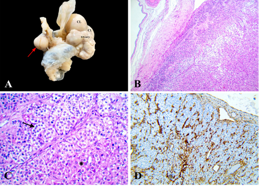
Adult Granulosa cell tumor, ovary, camel. (A) Firm grayish white nodule attached to the ovary by a long stalk (black arrow), Inset, hemorrhagic cut section. (B) Neoplastic cells arranged in solid sheets, supported by fine fibrovascular stroma, HE x 200. Inset, round to ovoid uniform nuclei with pale chromatin and coffee-bean-like nuclear groove, HE x 400. (C) Sarcomatous change characterized by pleomorphic cells with irregular, hyperchromatic, bizarre nuclei (arrowhead) showing variable mitosis, HE x 400. (D) Neoplastic cells expressed cytoplasmic positive reaction for vimentin, x 200.
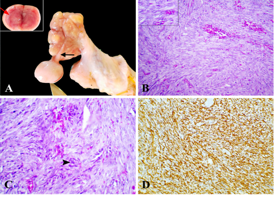
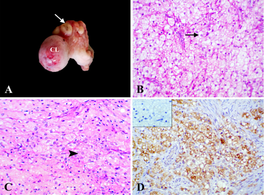
Interstitial cell tumor, ovary, camel. (A) Firm grayish white nodule (arrow) attached to mesovarium, CL; corpus luteum. (B) Encapsulated moderately cellular neoplasm, HE x 100. (C) Neoplastic cells arranged in solid sheets, cords (asterisk), and indistinct nests or acini (arrow) separated by fine fibrovascular stroma, HE x 400. (D) Neoplastic cells expressed cytoplasmic positive reaction for vimentin, x 200.

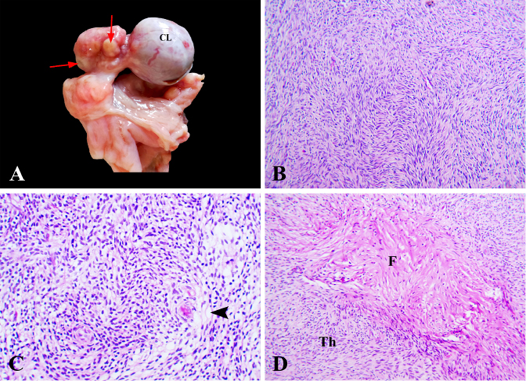
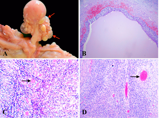
Thecoma, ovary, camel. (A) Small firm grayish white nodules (arrow), CL; corpus luteum. (B) Aggregates of streaming oval and spindle cells arranged in a diffuse pattern, HE x 200. (C) Neoplastic cells had pale vacuolated cytoplasm (arrowhead), HE x 400. (D) Fibrothecoma characterized by mixtures of fibroma (F) and thecoma (Th), HE x 200.

Steroid cell tumor, NOS, ovary, camel. (A) Firm grayish white to yellow nodules (arrow) CL; corpus luteum. (B) Neoplastic cells had marked amounts of foamy or clear cytoplasm (arrow), HE x 400. (C) Neoplastic cells had eosinophilic or yellowish-pigmented cytoplasm (arrowhead), HE x 400. (D) Neoplastic cells expressed cytoplasmic positive reaction for melan A, x 400. Inset, negative reaction for inhibin, x 400.

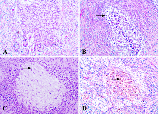
Granulose-theca cell tumor, ovary, camel. (A) Firm grayish-white nodules and numerous yellow cysts (arrow). (B) Macrofollicular pattern with hemorrhage and eosinophilic secretion, HE x 100. (C) Trabecular pattern with Call-Exner bodies (arrow), HE x 400. (D) Diffuse pattern with Call-Exner bodies (arrow), HE x 200.
Granulose-theca cell tumor, uncommon pattern, ovary, camel, HE x 400. (A) Solid follicular pattern without lumen. (B) Luteinized-granulosa (arrow) pattern. (C) Luteinized-thecoma (arrow) pattern. (D) Luteinized-pigmented pattern characterized by brownish granules (arrow).

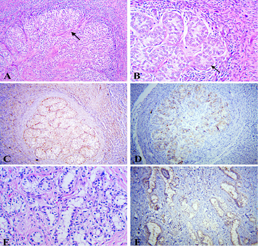
Granulose-theca cell tumor, ovary, camel. (A) Insular pattern with dense fibrous stroma (arrow), HE x 200. (B) Tubule-like (arrow) pattern, HE x 400. (C) Tubule-like pattern expressed strong diffuse positive reaction for vimentin, x 200. (D) Tubule-like pattern expressed moderate focal positive reaction for inhibin, x 200. (E) Sertoli-like pattern, HE x 400. (F) Sertoli-like pattern expressed cytoplasmic positive reaction for melan A, x 200.


