
Computerized Tomography, Radiological and Morphological Features on the Skull of Egyptian Owl (Bubo ascalaphus)


Computerized Tomography, Radiological and Morphological Features on the Skull of Egyptian Owl (Bubo ascalaphus)
Ramadan Sary, Asmaa M. Ibrahium*

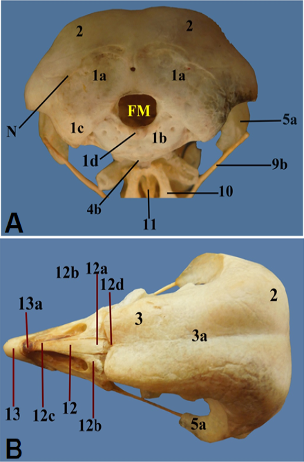
Figure 1:
A photograph showing; (A) Occipital region of cranium; (B) Dorsal aspect of cranium.

Figure 2:
A photograph showing; (A) Ventral aspect of cranium; (B) Quadrate bone.

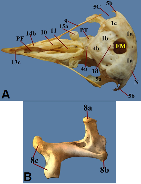
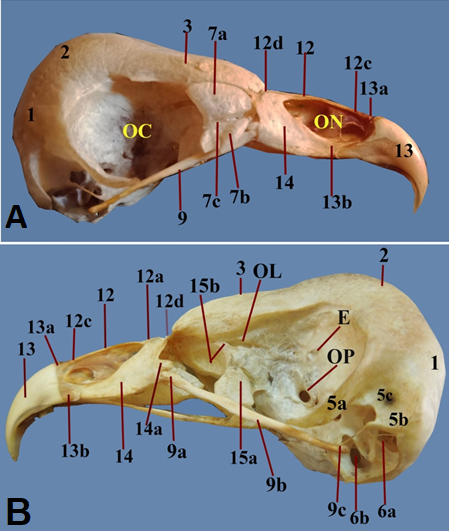
Figure 3:
A photograph showing lateral aspect of cranium.
(A) before removing lacrimal bone; (B) After removing lacrimal bone.
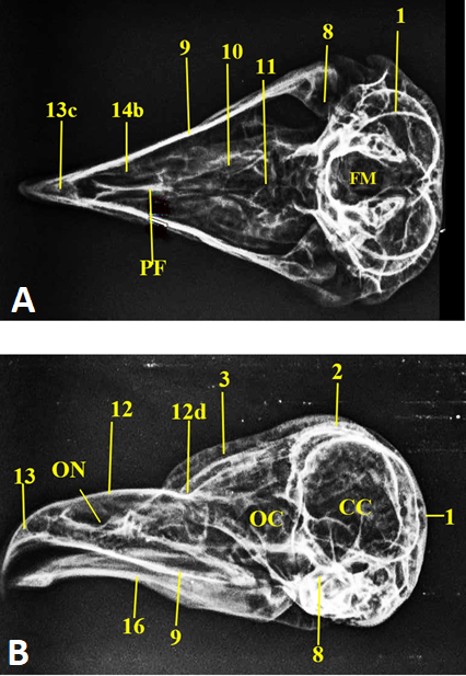
Figure 4:
A photograph showing X-ray image of whole skull; (A) Ventral view; (B) Lateral view.

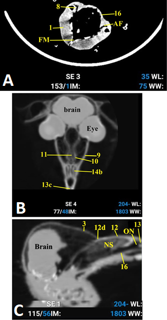
Figure 5:
A photograph showing CT image of whole skull. A: caudal view; B: Ventral view; C: Lateral view.
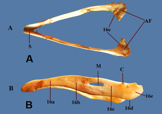
Figure 6:
photograph showing the mandible. A: Dorsal view; B: Lateral view.
1-Occipital bone, 1a-Supraoocipital part, 1b-Basioociptal part, 1c- Exo-oxcipital part, 1d-Occipital condoyle, 2-Parital bone, 3-Frontal bone, 3a- longitudinal fissure, 4- Sphenoid bone, 4a-Presphenoid, 4b-Basisphenoid, 5-Squamus part of temporal bone, 5a-orbital process, 5b-Zygomatic process, 5c-temporal fossa, 6- Ear capsule, 6a- Internal acoustic meatus, 6b- Tympanic cavity proper, 7- Lacrimal bone, 7a- dorsal lacrimal process, 7b-ventral lacrimal process, 7c- deep fissure, 8-Quadrate bone, 8a- Orbital process, 8b- Otic process, 8c- Articular process, 9- Zygomatic (Jugal) bone, 9a Jugal process , 9b- Proper jugal, 9c- Quadrojugal process,10- Palatine bone, 11-Vomer bone, 12- Nasal bone, 12a-Frontal process of Nasal, 12b- Maxillary process of Nasal, 12c- Premaxillary process of Nasal, 12d- Frontonasal hing 13- Inscisive bone (premaxillary bone), 13a- Frontal process of incisive, 13b- Maxillary process of incisive , 13c- Palatine process of incisive, 14-Maxillary bone, 14a-Zygomatic process, 14b- Palatine process of maxilla, 15-Ethmoid bone, 15a –vertical part of Ethmoid, 15b- Horizontal part of Ethmoid, 16- Mandible, 16a- Dental bone, 16b-splenial bone, 16c- supra angular bone, 16d-The angular bone, 16e-The articular bone. N: Nuchal crest, FM: Foramen Magnum, OP: Optic foramen, E: Ethmoid foramen, PF: Palatine fissure, PT: Pterygoid facet, M: Mandibular foramen, C: Coronoid process, S: Mandibular Symphysis, AF: Articular facet for quadrate bone, OL: olfactory foramen.OC: Orbital Cavity, ON: Osseous opening of nostril, NS: Nasal Septum, CC: Cranial cavity.

August 2022
Vol. 10, Iss. 8, Pages 1659-1886
