
Cyto-morphological-Changes-in-Exfoliated-Vaginal-Cells-and-Thermal-Rhythms-of-Red-Sokoto-does-during-the-Oestrous-cycle


Cyto-morphological-Changes-in-Exfoliated-Vaginal-Cells-and-Thermal-Rhythms-of-Red-Sokoto-does-during-the-Oestrous-cycle
Umar M. Bello1*, Samuel A. Ojo1, Abdurrahman Ghaji1, Ambrose A. Voh (Jr)2, Muazu N. Bappah3, Casmir O. Igbokwe4

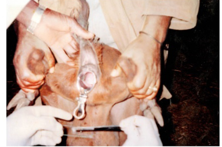
Exfoliated vaginal smear taken from the anterior vagina (5cm caudad to cervical opening) of Red Sokoto doe, using a self-retaining bivalved (BicuscoTM) speculum and with the aid of cytobrushTM, which then smeared onto a clean fat-free labelled micro glass slide.

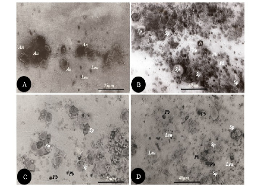
Photomicrographs of exfoliated vaginal cells smear taken during various stages of the estrous cycle A) Proestrus phase- showing several polyhedral superficial nucleated cells (An), single or in clusters and containing dark (glycogen) granules. Leucocytes (Leu) are seen but very scanty. B) Oestrus phase- Note the characteristic highly cellular nature of smear which is deeply basophilic stained. Superficial nucleated cells (Sp) are abundant, few parabasal (Pb) and Leucocytes (Leu) are seen. C) Metestrus phase- Note few superficial cells (polygonal in outline) and parabasal (Pb) cells, Leucocytes (Leu) begin to appear in this phase. D) Diestrus phase- Note the abundant leucocytes (Leu) i.e marked leukocytosis which is predominantly seen in the smear, also few superficial nucleated cells (Sp) are seen. Modified Papanicolaou stain.
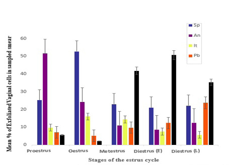
Percentage of epithelial vaginal cells (mean ± SEM) of the vaginal smears from Red Sokoto does (n=15) PGF2induced estrus at different stages of the Oestrus cycle. Legend: Sp- Superficial nucleated cells; An- Anucleated superficial; It- Intermediate cells; Pb - Parabasal; Leu- Leucocytes.

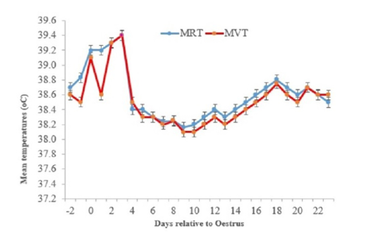
The mean (± SEM) of rectal and vaginal temperature values of Red Sokoto does (n=15) PGF2α- induced estrus at various stages of the Oestrus cycle. Legend: Mean Rectal temperature (MRT), Mean Vaginal temperature (MVT).


