
Dermatophytosis in Clinically Infected Cats: Diagnoses and Efficacy Therapy


Dermatophytosis in Clinically Infected Cats: Diagnoses and Efficacy Therapy
Alsi Dara Paryuni1, Soedarmanto Indarjulianto2, Tri Untari3 and Sitarina Widyarini4*
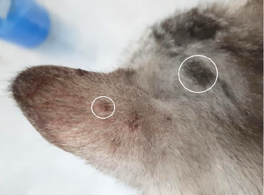
Figure 1:
Lesions from dermatophyte infection in body part of cat with alopecia, erythema, and scale in cat dermatophytosis (white ring).
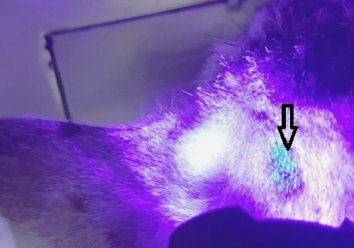
Figure 2:
Ear of cat with fluorescence (apple blue-green color), under Wood’s lamp examination (black arrow).

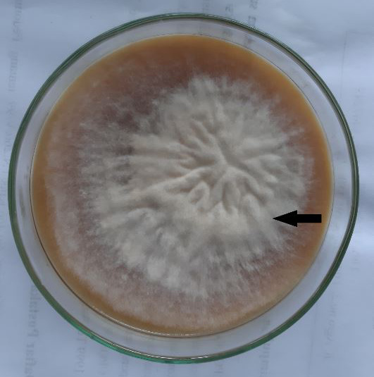
Figure 3:
Fungal colony of Microsporum canis; Cottony, white to buff in colour; with increasing age becomes orange-brown (black arrow).
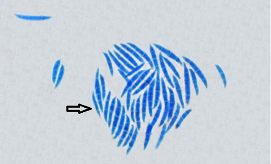
Figure 4:
Microscopic structure of Microsporum canis with Lactophenol cotton blue stain (black arrow).

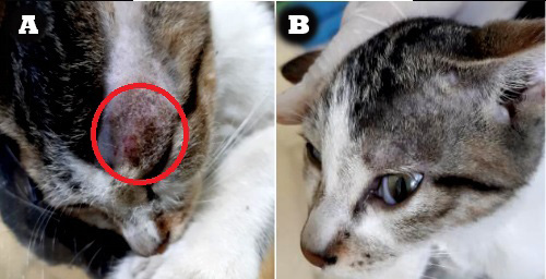
Figure 5:
Macroscopic lesions progress (decrease in severity) on treatment with 2% Ketoconazole cream: A. Before treatment (red circle), B. The last day (day 21st) of treatment.

August 2022
Vol. 10, Iss. 8, Pages 1659-1886
