

Evaluation of Inflammatory Cytokine IL-17 and Immunohistochemical Analysis of Cirrhotic Liver from HCV Patients


Evaluation of Inflammatory Cytokine IL-17 and Immunohistochemical Analysis of Cirrhotic Liver from HCV Patients
Shafi Muhammad1, Bibi Nazia Murtaza2, Aftab Ahmad1, Muhammad Shafiq3, Nurul Kabir4 and Hamid Ali1*
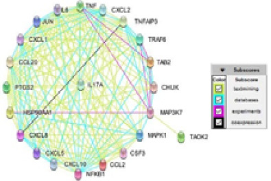
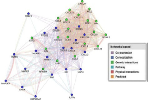
IL-17A gene interaction network using the STRING database.
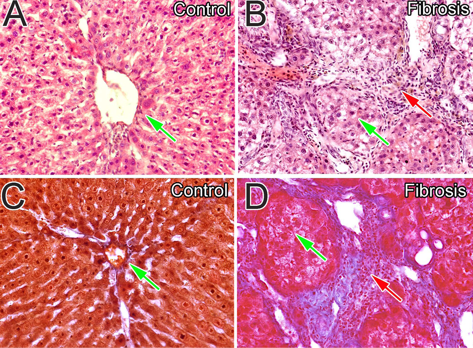
H&E and Masson’s trichrome staining of liver tissues of HCV infected patients. A, Stained with H/E, the control group with no sign of any abnormality; B, H/E shows cords of fibrosis with mixed infiltration of inflammatory cells; C, Masson’s trichrome staining normal control group with distinct shaped nuclei of hepatocytes and their ar-rangement in cords; D, Masson’s trichrome staining showing extensive fibrosis. The shapes of hepatic lobes and lobules were distorted in the fibrosis group.
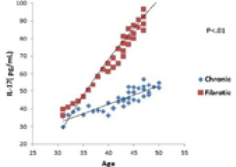
The correlation of IL-17A serum levels with age in chronic vs. fibrotic patients.

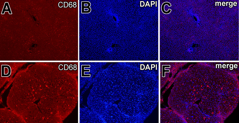
Localization and expression of macrophages in normal and fibrotic liver. Reduced the number of CD68+ cells: Double fluorescence for CD68 and DAPI in normal liver (A, B, C) and in the fibrotic liver showed numerous CD68+ cells along the sinusoids around the central vein (D, E, F).

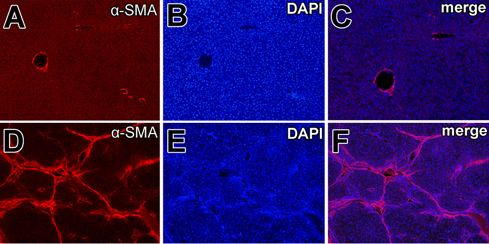
Immunohistochemistry of alpha SMA in the liver of HCV positive patients. Normal control group with alpha SMA, DAPI and marge (A, B, C) While in the cirrhotic group there is huge expression of alpha SMA making bridging fibrosis (D, E, F).


