

Hepatoprotective Potential of Mesembryanthemum forsskalii Fruits Extract Against Carbon Tetrachloride-Induced Liver Toxicity in Mice


Hepatoprotective Potential of Mesembryanthemum forsskalii Fruits Extract Against Carbon Tetrachloride-Induced Liver Toxicity in Mice
Usama Mahalel1, Barakat M. Alrashdi1, Ibrahim Abdel-Farid1, Sabry El-Naggar2, Mohamed Hassan3, Hassan Elgebaly1 and Diaa Massoud1,4*

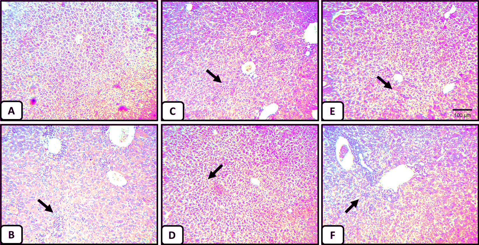
Histopathology of liver tissues. (A) Liver section of normal negative control mice (vehicle) shows central vein surrounded by hepatic cord of cells (normal architecture), (B) liver section of CCl4-treated mice showing massive fatty changes, focal central vein congestion, a variety of cavitations and necrosis in hepatocytes with inflammation, and loss of cellular boundaries (indicated by arrow), (C) liver section of mice treated with 100mg/kg showing normal liver architecture (magnification 5X), (D) liver section of mice treated with the 500mg/kg showing normal liver architecture, (E) liver section of mice treated with CCl4 and 100mg/kg of M. forsskalii showing absence of cavitations, necrosis, and inflammatory cells, and regeneration of hepatocytes around central vein toward near normal liver architecture but slight congestion in central vein (indicated by arrow), and (F) liver section of mice treated with CCl4 and 500 mg/kg of M. forsskalii showing mild central vein congestion (indicated by arrow), ballooning, and necrosis with sinusoidal dilatation H and E x 50.

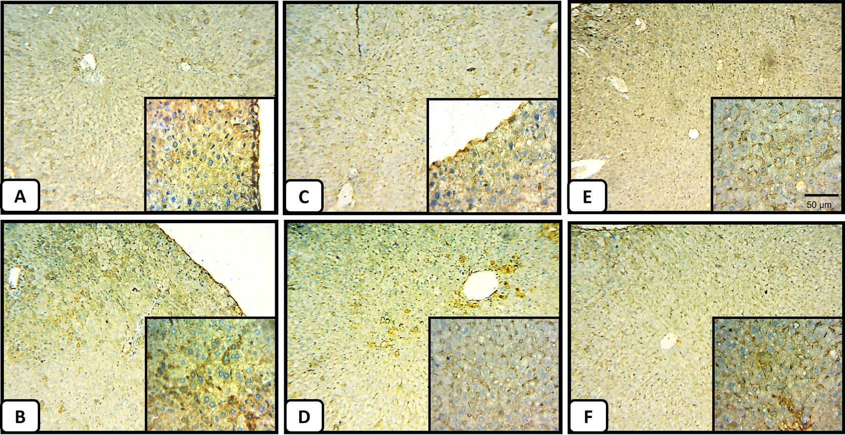
Immunohistochemistry of P53 in the liver tissues. (A) Section from a negative control mice liver shows the normal pattern of P53 with mild staining. (B) The liver section obtained from CCl4-intoxicated mice shows extensive staining (indicated by brown color) for P53. (C) and (D) a liver section from normal mice treated at low and high doses, respectively, showing a non-appreciated difference in P53 staining pattern (magnification, x 50 for the main photo and x 100 for the hyper-focused region in the small box at the left bottom of each panel). (E) and (F) Liver tissue sections prepared from the 100 mg/kg and 500mg/kg of M. forsskalii after CCl4 treatment, respectively, show less P53 staining compared to (B), however (E) shows a closer staining pattern to the normal liver (A).

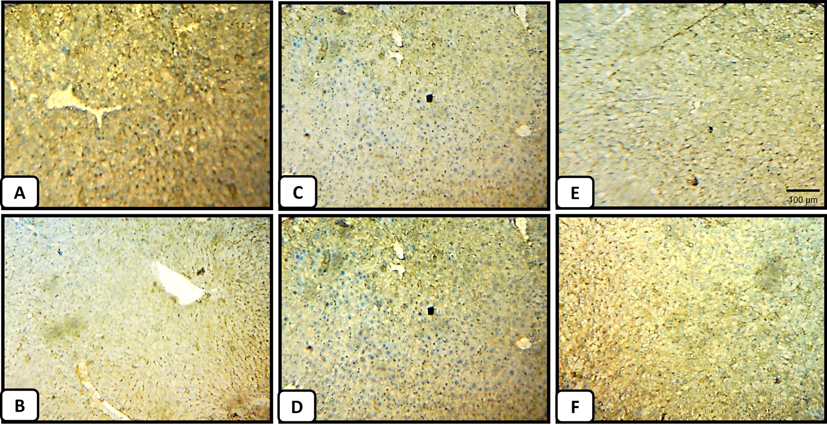
Immunohistochemistry of the cytoprotective molecule, Bcl-2, in the liver tissues. (A) A representative section from a negative control mice liver shows a normal pattern of Bcl-2 with mild staining. (B) The liver section obtained from CCl4-intoxicated mice shows faint staining (indicated by brown color) for Bcl-2. (C) Liver section from normal mice treated with M. forsskalii extract at a low dose shows a detectable reduction in the expression of Bcl-2 in the normal liver when introduced at a low dose (Magnification, x 50), (D) A representative liver section from normal mice treated at high dose shows mild staining ability by Bcl-2, indicating a very limited effect of M. forsskalii extract on Bcl-2 expression when administered at high dose, (E) and (F) Liver tissue sections prepared from the 100 mg/kg and 500 mg/kg of M. forsskalii after CCl4 treatment groups, respectively, showing the relative restoration of the Bcl-2 staining compared to (B), however (F) shows closer staining pattern to the normal liver (A).


