
Histopathological Examination of the Possibility of a Purrfect Neuro Inflammatory Storm Leading to Choroid Blindness and Death Following Traumatic Brain Injury in Red Kangaroo (Osphranter rufus)


Histopathological Examination of the Possibility of a Purrfect Neuro Inflammatory Storm Leading to Choroid Blindness and Death Following Traumatic Brain Injury in Red Kangaroo (Osphranter rufus)
Peyman Mohammadzadeh1*, Aida Rasuli2, Bahman Noroozi Gorgani2, Sajjad Mohammadi3

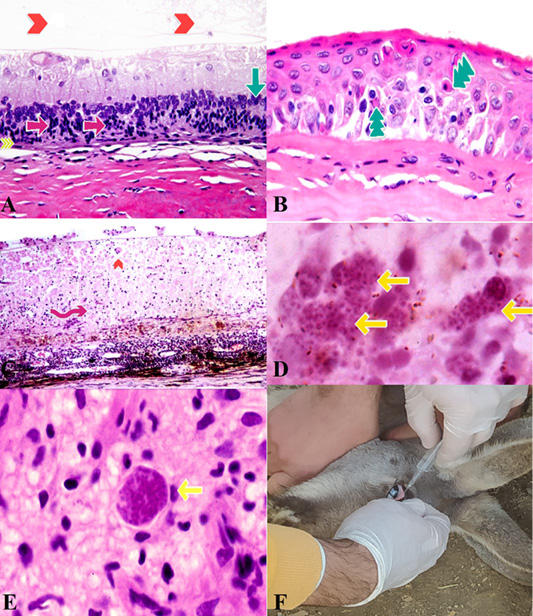
Eye, change in Traumatic Brain Injury. A: Mild degeneration of Retina, including loss of cone and rod photoreceptor processes, (yellow arrowheads), unicellular necrosis (single-cell) in the outer part of nuclear layer photoreceptor cells, (Pink Arrows), Disorganization and hypo cellularity of the outer and inner parts of the nuclear layers, (Green Arrow) and narrowing and absence of the plexiform layers. (redhead arrow). (H & E, x800). B: Cornea ,Necrosis. Hypereosinophilic and shrunken necrotic cells, (Green Arrow) (H & E, x800). C: overlying necrosis of retina, (Pink Arrow) and presence of a large Toxoplasma gondii tissue pseudocyst on the surface. (redhead arrow) (H & E, x600). D, E: Toxoplasma gondii cyst-containing discrete granule-type bradyzoites (yellow arrows) (H & E, x800). F: Intraocular injection in another patient kangaroo.
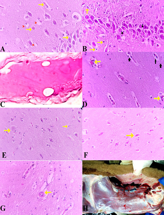
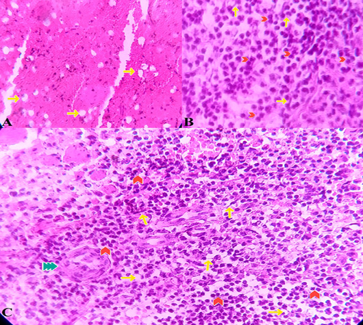
Brain changes in Traumatic Brain Injury. The early changes reflect alterations in ion gradients accompanying shifts in the perikaryon (Soma) and cause cloudy swelling. A: Swelling of the endoplasmic reticulum appears as a gap (cleft) in the periphery of the soma (Yellow arrows). This is different from crystalline inclusions and should not be mistaken. Astrocyte takes up Potassium + and the cytoplasm becomes pale due to intracellular fluid accumulation (red arrowhead) (H&E, x600). B: In response to brain-contusions injuries, neurons appear dark and pyknotic. (yellow arrows) (H&E, x200). astrocytes as well as Surveillant microglia activated to produce extra inflammatory mediators (Black Arrows). C: due to cellular edema and sufficient concentration of swollen neurons, some regions appear pale. (H&E, x12.5). D: necrotic neurons have a uniform eosinophilic cytoplasm. (yellow arrows). highe proportion of activated microglia and astrogliosis (Black Arrows) (H&E, x600). E: Some neurons become ghost cells without nucleus staining and a barely visible cell architecture (yellow arrows). (H&E, x600). F: rare eosinophilic neurons (yellow arrows) (H&E, x600). G: Toxoplasma gondii tissue cysts in the cerebral cortex (yellow arrows). (H&E, x600). H: Traumatic myositis in Cleidomastoid, Sternomastoid, Sternothyroid, Sternohyoid and Trapezius Muscles and sever damage to jugular vein, linguofacial vein, maxillary vein and caudal auricular vein, great auricular nerve lead to ruptures and hemorrhage.

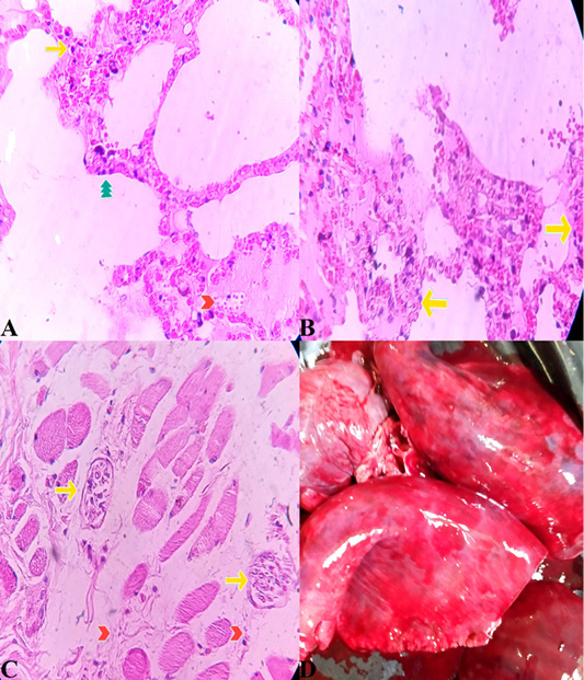
Lung changes in Traumatic Brain Injury. A: neutrophil infiltration (yellow arrow) and proliferation in the alveolar and interstitial space, thickening of the alveolar septum, (green arrow) as well as edema and alveolar hemorrhage (redhead arrow). (H&E, x600). B: destruction of type II alveolar cells, which leads to disruption of normal fluid transport. (yellow arrow). D: Toxoplasma gondii cyst-containing discrete granule-type bradyzoites (yellow arrows) and free tachyzoites (red arrowheads) (H&E, x600). C: Macroscopic view of Lung tissue.

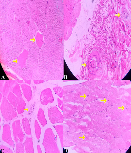
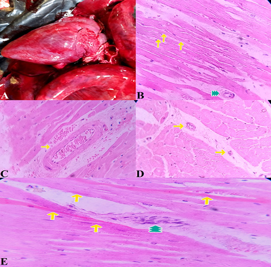
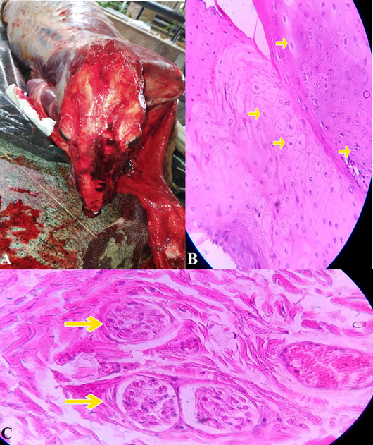
Muscle spino-acromio-clavodeltoideus. changes in Traumatic Brain Injury. A) Degenerated fibers, (yellow arrow) (H&E, x600). B, C: Toxoplasma gondii cyst-containing discrete granule-type bradyzoites (yellow arrows) (H&E, x600). D: Central nucleation can be seen in the injured M. infraspinatus (H&E, x600).

Spleen changes in Traumatic Brain Injury. A: internuclear bridges (yellow arrows) and binucleated erythroid precursors (redhead arrow) were readily recognized (H&E, x600). B: severe depletion of white pulp lymphatic tissue (yellow arrows) (H&E, x600). C: Toxoplasma gondii cyst-containing discrete granule-type bradyzoites (Green arrow) internuclear bridges (yellow arrows) and binucleated erythroid precursors (redhead arrow) (H&E, x600).

Heart changes in Traumatic Brain Injury. A: Macroscopic view of heart tissue. B: Degeneration and Atrophy of myocardial fibers (yellow arrows) Toxoplasma gondii cyst-containing discrete granule-type bradyzoites (Green arrow) (H&E, x600). C: Congestion and hemorrhage of cardiac tissue(H&E, x600). D: Toxoplasma gondii cyst-containing discrete granule-type bradyzoites (yellow arrow) (H&E, x600). E: Hypereosinophilic bundles(yellow arrows) and leucocyte infiltration(Green arrow) (H&E, x600).

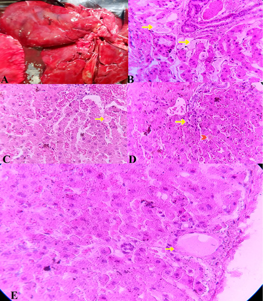
Liver changes in Traumatic Brain Injury. apoptosis, edema, and inflammation in hepatic tissue were increased. A: Macroscopic view of Liver tissue. B: Focal points of edematous fluid accumulation in the space between hepatocytes (yellow arrow) (H&E, x600). C: Rare apoptotic cell (yellow arrow) (H&E, x600). D: Infiltration of inflammatory cells (yellow arrow) Rare apoptotic cell (Red Head Arrow) (H&E, x600). E: Focal accumulation of edema (yellow arrows) (H&E, x600).

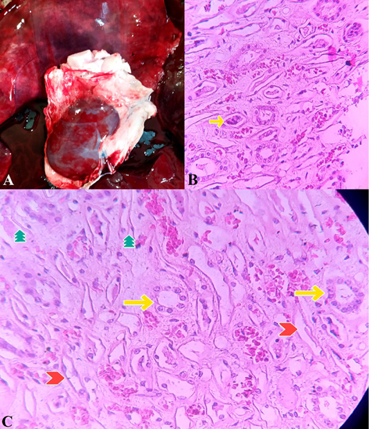
Kidney changes in Traumatic Brain Injury. A: Macroscopic view of Kidney tissue. B: Toxoplasma gondii cyst-containing discrete granule-type bradyzoites (yellow arrow) (H&E, x600). C: Damaged proximal cell tubules demonstrating epithelial thinning (Green arrows), epithelial blebs (Red Head arrows), and proximal tubular dilation (yellow arrows) (H&E, x600).

Tunge Muscle changes in Traumatic Brain Injury. A: Rupture of Levator nasolabialis, Levator labii maxiltaris, Temporalis, Masseter and Buccinator Muscles due to severe physical trauma that resulted in severe injury to Communicating branch of auriculotemporal nerve, transverse facial vein one facial artery and facial vein that lead to severe hemorrhage. B: hypoxic death cells appeared to be swollen and the cell membrane, as well as the nuclear membrane, was discernible. (yellow arrow) (H&E, x600). C: Toxoplasma gondii cyst-containing discrete granule-type bradyzoites (yellow arrows) (H&E, x600).

