
Improvements in Some Physiological and Histologic Aspects in the Rat Model When Lipoic Acid is Combined with Salbutamol


Improvements in Some Physiological and Histologic Aspects in the Rat Model When Lipoic Acid is Combined with Salbutamol
Huda K. Khassaf1*, Zainab A.H. Al-Mousawi1, Sawsan A. Ali2, Anwar Nather Seiwan3
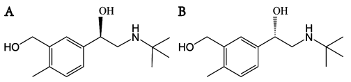
Chemical structure of (A) (R)-salbutamol and (B) (S)-salbutamol.

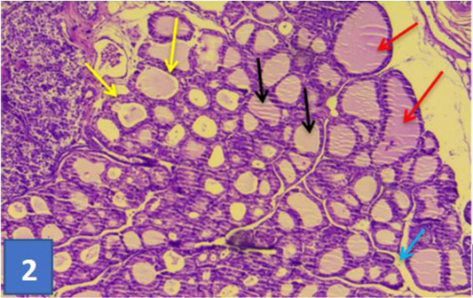
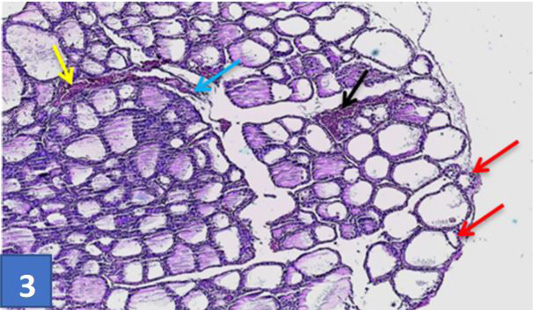
Section of thyroid gland of control group: showing normal histological appearance of follicles (red darts) of different size lining with simple epithelial (yellow darts), colloid substance (black darts), this connective tissue septa separated gland lobules (blue dart). (H & E) stain (10X).

Section of thyroid gland treated by SAL: showing atrophied with shrinking follicles (red darts), congested large blood vessel (yellow dart), and hyperplastic changes (black dart) thickness of connective tissue separated the thyroid lobules (blue dart) (H & E) stain (10X).

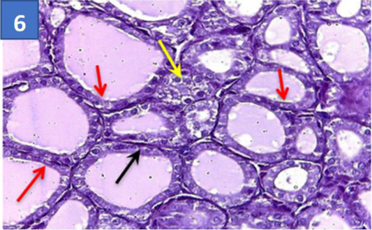
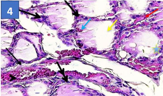
Section of thyroid gland treated by SAL: Showing thickness of tissue seprated of the lobules (black darts)moderete depletation of para follicular cell (thick arrows), scanty colloid (yellow dart) hypermia between thyroid follicles(red dart),and between thyrocyte (blue dart), infiltrated of inflmmatory cell around some follicles (double arrow), hyperplastic capillaries (arrow head) and intrafolliculor hyperplasia (blue star) (40X).
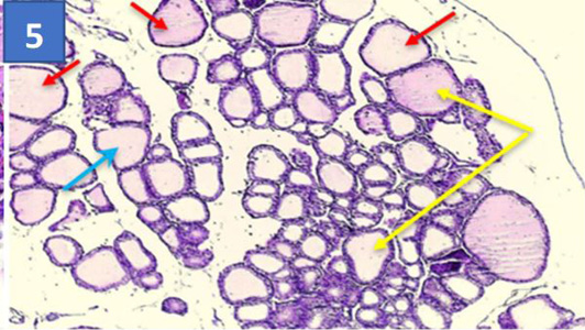
Section of thyroid gland treated by SAL+ALA: showing more large regular follicles thyroid (red darts) follicles hyperplastic intrafollicular connective tissue (blue dart), some large follicle with partially amount of tissue (yellow darts) (H & E) stain (10X).
Section of thyroid gland treated by SAL+ALA: Showing more improvement thyroid follicle wall lined with typical cuboid epithelial cells (red darts), with clear parafollicular cells (yellow darts), thin septa of collagen fibers between thyrocytes and follicles (black dart) (H & E) stain(40X).

