
Male Reproductive System Cytotoxicity and Immunotoxicity Evaluation Following Folcord Exposure


Male Reproductive System Cytotoxicity and Immunotoxicity Evaluation Following Folcord Exposure
Salema Lafta Hassan1*, Taghred Jabbar Humadai1, Sabrin Ibraheem Mohsin2
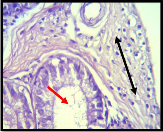
A histopathological slice of the first group’s epididymes at 4 weeks demonstrates vacuolated cells, poorly differentiated spermatogenic cells (red arrow), and interstitial edema. Interstial tissue with inflammatory cell infiltration (black arrow) (H & E stain X 40).
A histopathological slice of the first group’s testes at 4 weeks reveals severe vacuolations of the seminiferous tubular epaithelium with the lack of spermatogenic cells (black arrow) (H & E stain X 40).

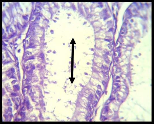
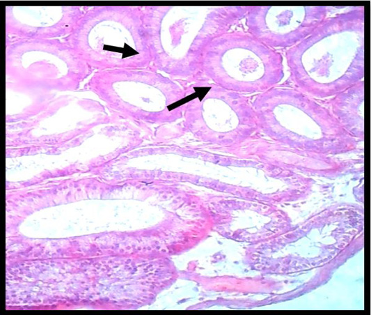
A histopathological slice of the first group’s epididymis at 4 weeks demonstrates that there is no epididymal sperm reserve in the tubules. The lumen included homogeneous material and cellular detritus. (black arrow) (H & E stain X 10).

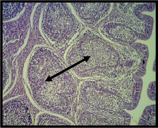
Histopathological slice of the first group’s epididymis at 4 weeks demonstrates granuloma development (black arrow) (H & E stain X 40).
A histopathological slice of the second group’s testes after 4 weeks indicates considerable deterioration and the absence of testes sperm (black arrow) (H&E stain X 10).

Sertoli cells are somewhat vacuolated in the second group’s testes at 4 weeks, and the creation of big cells does not continue with the spermatogenic cell layer (H & E stain X 40).

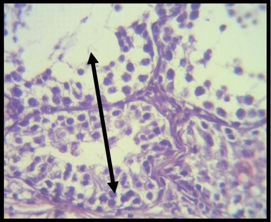
Histopathological slice of the second group’s testes at 4 weeks shows inflammatory cell aggregation in moderately vacuolated interstial tissue, and the layer of spermatogenic cells is discontinuous. (black arrow) (H & E stain X 40).

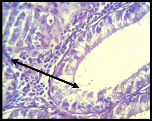
Histopathological slice of the second group’s testes at 4 weeks exhibits hyperplasia of clear cells that are strongly vacuolated, homogeneous material, and cellular debris (black arrow) in the lumen (H & E stain X 40).
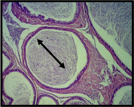
Histopathological section of testes at 4 weeks of 3rd group shows at various phases of spermatogenesis, seminiferous tubules form, and the Leydig cells and mature spermatozoa fill the lumen. (black arrow) (H&E stainX10).

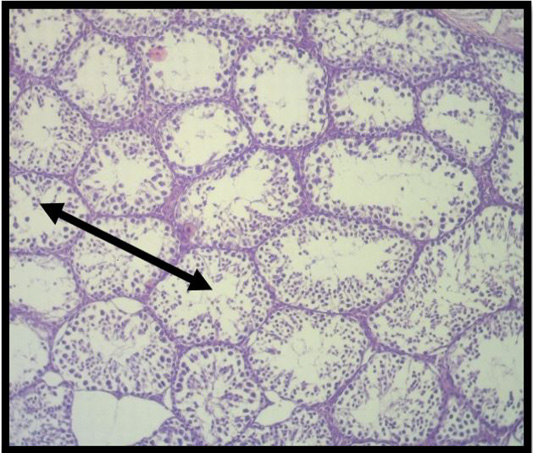
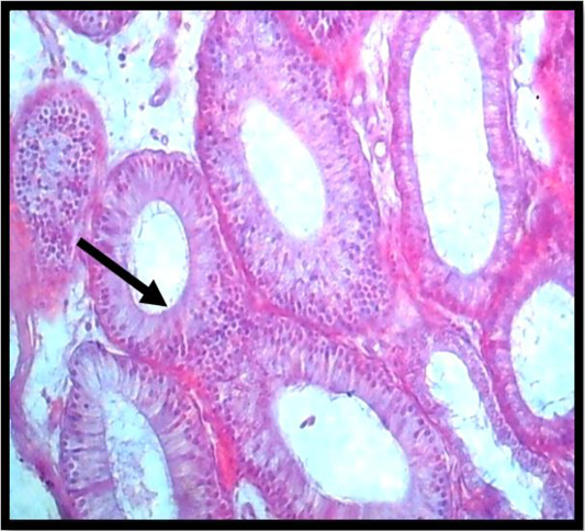
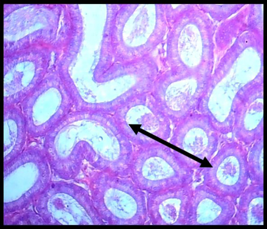
A histopathological slice of the third group’s testes at 4 weeks demonstrates full epididymal sperm reserve in the tubules (black arrow) (H & E stain X 10).

