Nano-Carbonated Hydroxyapatite from Bovine Bone Combined With Platelet-Rich Plasma Accelerates Fracture Healing in Rattus norvegicus Models
Nano-Carbonated Hydroxyapatite from Bovine Bone Combined With Platelet-Rich Plasma Accelerates Fracture Healing in Rattus norvegicus Models
Zahra Jamilah Sabrina1, Adrian Pearl Gunawan2, Beryl Reinaldo Chandra2, Ilham Pangestu Harwoko3, Johannes Marulitua Nainggolan4,Widi Nugroho1*
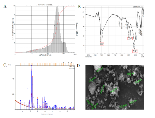
Characteristics of CHA; A. Particle size distribution of micro-CHA analyzed using PSA; the mean size of the particles is 39.98 μm (SD: 4.21 - 80.97 μm); B. The FTIR analysis of micro-CHA shows functional groups of OH-, CO32- and PO43-; C. The XRD analysis of nano-CHA shows the prominent diffraction peaks formed at the diffraction angle of 31.74°; D. The particle size distribution of nano-CHA analyzed using SEM at 10.000x magnification; the particles were of spherical shapes, a few particles were joining together to form agglomerates with the size of agglomerates ranging 82.68 - 153 nm.
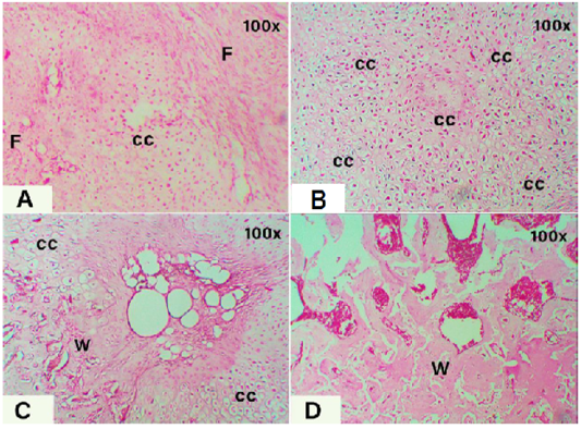
Sections of fractured tibial bone tissues at 21 days post-treatments. A. control group, showing the formation of fibrocartilage tissue by chondrocyte cells (cc) in between the fibrous tissue (F) which was developed earlier; B. Micro-CHA group showing the complete formation of fibrocartilage tissue (Fc) indicates the late phase of soft callus formation; C. Micro-CHA+PRP group, showing the domination of fibrocartilage tissue and the formation of a small proportion of woven bone tissue (W) indicating a hard callus formation; D. Nano-CHA+PRP group, showing the formation of new bone matrix in the fractured area indicated by the domination of woven bone and fusion into lamellar bone. H&E staining, 100x magnification.


