Potential Pathological Effects After Repeated Exposure to Tetrodotoxin in Reproductive System of Albino Male Rats
Taghreed Jabbar Humadai*, Bushra Ibrahim AL-Kaisei
Department of Pathology and Poultry Diseases, College of Veterinary Medicine, University of Baghdad, Baghdad, Iraq.
*Correspondence | Taghreed Jabbar Humadai, Department of Pathology and Poultry Diseases, College of Veterinary Medicine, University of Baghdad, Baghdad, Iraq; Email: tagreed.j@covm.uobaghdad.edu.iq
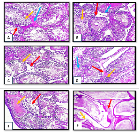
Figure 1:
Histopathological section of testes and epididymis tissue from tetrodotoxin treated group (0.5 µg/kg.bw) of male albino rats (A) sever degenerated changes of spermatogonia cells (red arrow) with irregular appearance of spermatid (yellow arrow) with presence of edematous substance between degenerated seminiferous tubules. (B) obvious disruption of seminiferous tubules (red arrow) with necrotic finding of some tubules (yellow arrow) with absence of leydig cells (blue arrow). (C) complete loss of leydig cells (red arrow) with evidence of hypospematogensis (yellow arrow). (D) intertubler edema (red arrow) with mild mononuclear cells infiltration (yellow arrow) with necrotic of immature spermatid (blue arrow). complete necrosis of spermatogonial cells (red arrow) with obvious subcapsulear vascular congestion and dilation (yellow arrow). (E) complete necrosis of spermatogonial cells (red arrow) with obvious subcapsulear vascular congestion and dilation (yellow arrow). H&E, x10. (F) marked irregularity of epididymal tubules (red arrow) with obvious atrophy of some tubules (yellow arrow). H&E, x40
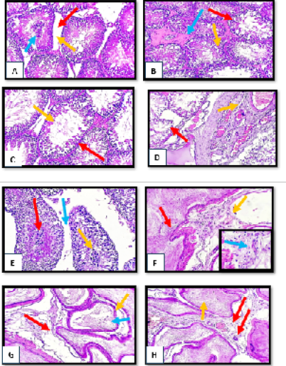
Figure 2:
Histopathological section of testes and epididymis tissue from tetrodotoxin treated group (1µg/kg.bw) of male albino rats (A) disruption of seminiferous tubules (red arrow) with absence of leydig cells (yellow arrow) with central clamping of necrotic spermatogonia (blue arrow).(B) sever degenerative changes of spermatogonia cells (red arrow) with irregular appearance of spermatid (yellow arrow) with presence of edematous substance between degenerated seminiferous tubules (blue arrow).(C) marked reduction of spermatogonia cells lining (red arrow) with sperm irregularity (yellow arrow).(D) disorganized irregular seminiferous tubules (red arrow) with subcapsular dilation and congestion (yellow arrow).(E) clumping of degenerated spermatocyte (red arrow) with necrotic sperms (yellow arrow) with obvious widening of interlobular septa (blue arrow).(F) focal hyperplasia of epididymal lining (red arrow) with blood vessels congestion (yellow arrow) with sever mononuclear cells infiltration in the vascular connective tissue (blue arrow) (H&E stain, X10, inset X40).(G) moderate widening of some epididymal tubules (red arrow) with giant tubules appearance (yellow arrow) with slight degeneration of mature sperms (blue arrow) (H&E stain, X10, inset X40).(I) fibrovascular tissue with newly form capillaries between epididymal tubules (red arrow) with sperms stasis (yellow arrow). (H&E stain, X10).
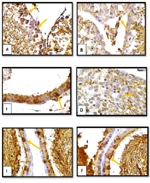
Figure 3:
Immunohistochemistry section of testes and epididymis tissue from tetrodotoxin treated group (A,B,C) (0.5µg/kg.bw),(D,E,F) (1µg/kg.bw) of male albino rats (A),(B) moderate immune positive cells for IL 1 antibody (arrow)mainly in sertoli cells and spermatogonia.(C) numerous immune positive cells for IL 1 antibody (arrow)in the epididymal epithelial cells lining.(D) numerous immune positive cells for IL 1 antibody (arrow) in spermatocyte. (E) moderate immune positive cells for IL1 antibody (arrow) in epididymal epithelial cells lining.(F) numerous immune positive cells for IL 1 antibody (arrow) in epididymal epithelial cells lining. (DAB- chromogen, X40).
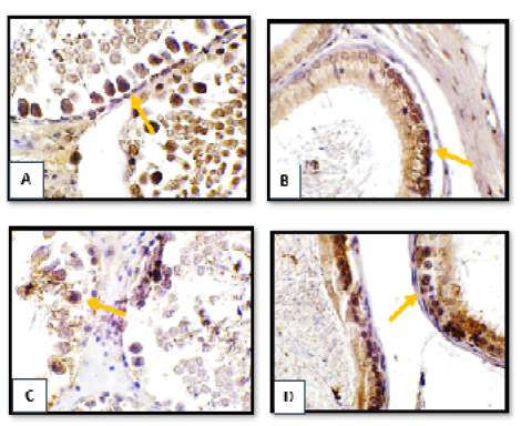
Figure 4:
Immunohistochemistry section of testes and epididymis tissue from tetrodotoxin treated group (A, B) (0.5µg/kg.bw), (C, D) (1µg/kg.bw) of male albino rats (A) numerous immune positive cells for TNFα antibody (arrow) in sertoli cells and spermatogonia. (B) moderate immune positive cells for TNFα antibody (arrow) epididymal epithelial cells lining. (C) mild immune positive cells for TNFα antibody (arrow) mainly in spermatogonia. (D) numerous immune positive cells for TNFα antibody (arrow) in epididymal epithelial cells lining. (DAB- chromogen, X40).


