

The Potential Role of Bone Marrow Derived Mesenchymal Stem Cells Conditioned Medium in the Suppression of Hepatocellular Carcinoma In Vitro: Modulation of Apoptosis


The Potential Role of Bone Marrow Derived Mesenchymal Stem Cells Conditioned Medium in the Suppression of Hepatocellular Carcinoma In Vitro: Modulation of Apoptosis
Mahmoud Abdelhady Metwally1*, Mona Abdelftah Ali1, Farouk Abdelmohdy1, Mohamed Kassab1, Tarek Kamal Abouzed2 and Khalil Fathy Abou-Easa1
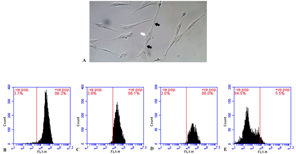
The identification of rat Bone marrow mesenchymal stem cell (BMMSCs) by microscopy and flowcytometry. The data shows a microscopic image (A) of BMMSCs, taken after 4 days in culture. The cells appear spindle-shaped, with a central cell body (white arrow) and cytoplasmic processes in opposite directions (black arrows). The data also shows flowcytometric identification of BMMSCs, revealing positive expression of specific surface markers: CD44 (B), CD90 (C), and CD105 (D) and negative expression for hemopoietic and endothelial surface marker CD34 (E).
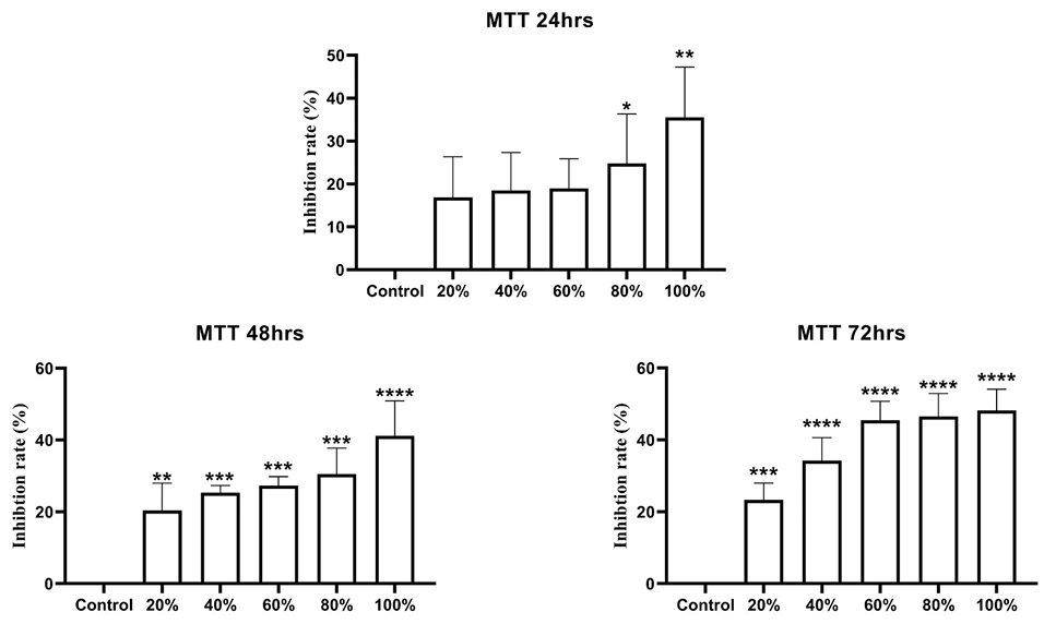
The inhibitory effect of Bone marrow mesenchymal stem cells-conditioned medium (BMMSCs-CM) on the hepatocellular carcinoma cell line HepG2 by MTT assay. The data shows the inhibition rate (%) of untreated HepG2 cell group (control) and treated HepG2 cell groups with different concentrations of BMMSC-CM (20%-100%) over 24, 48, and 72 h. Values are expressed as mean±SD, (n= 3/Time group), each concentration group/time was compared to the control group. (*P<0.0, ** P <0.01, *** P <0.001, **** P <0.0001).

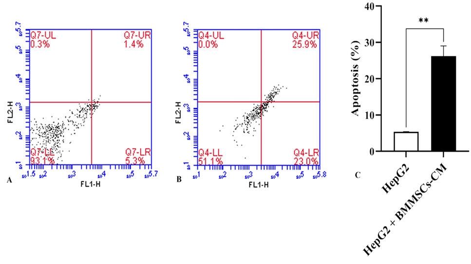
The apoptotic effect of Bone marrow mesenchymal stem cells-conditioned medium (BMMSCs-CM) on the hepatocellular carcinoma cell line HepG2 using Annexin-Propidium Iodide (PI) stains by flowcytometry. The data shows Annexin-PI measurements of untreated HepG2 cell group (control) (A) and treated HepG2 cell groups with 100% BMMSC-CM for 72 h (B), where the x-axis represents Annexin V-Fluorescein Isothiocyanate (FITC) saining and the y-axis represent PI staining, and the statistical analysis (C). Values are expressed as mean±SD, (n= 3), the treated group (HepG2+BMMSC-CM) was compared to the control group (HepG2). (** P<0.01).

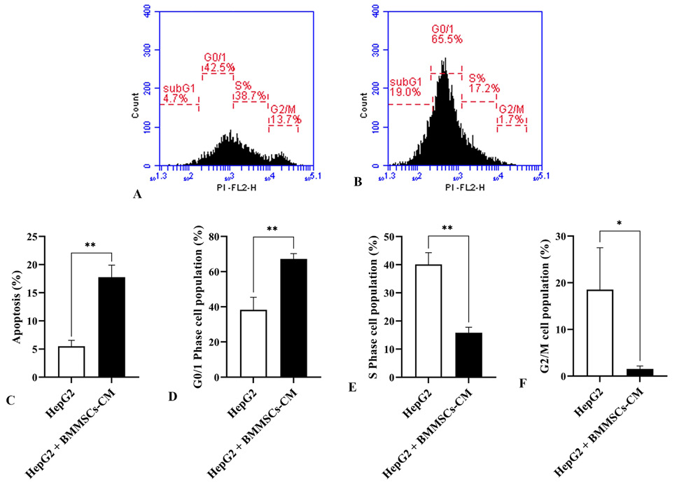
The impact of bone marrow mesenchymal stem cells-conditioned medium (BMMSCs-CM) on the hepatocellular carcinoma cell line HepG2 cell cycle by flowcytometry. The data shows the cell cycle of untreated HepG2 cell group (control) (A) and treated HepG2 cell groups with 100% BMMSC-CM for 72 h (B), The data shows the statistical analysis for each phase of the cell cycle (SubG1 phase (apoptosis)(C), G0/1 phase (D), S phase (E), and G2/M phase (F). Values are expressed as mean±SD, (n=3), the treated group (HepG2=BMMSC-CM) was compared to the control group (HepG2). (*P<0.05, **P<0.01).
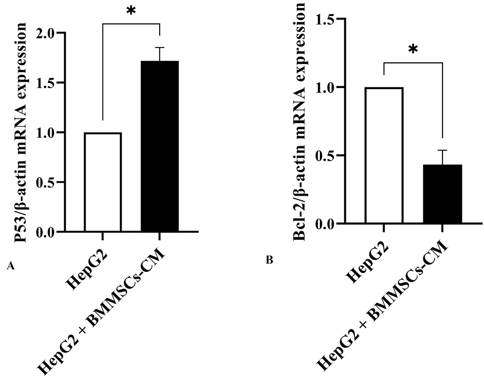
The influence of bone marrow mesenchymal stem cells-conditioned medium (BMMSCs-CM) on the hepatocellular carcinoma cell line HepG2 mRNA expression pf P53 gene (A) and Bcl-2 gene (B) by RT-PCR. The data shows the untreated HepG2 cell group (control) and treated HepG2 cell groups with 100% BMMSC-CM for 72 h. Β-actin was used as the house-keeping gene. Values are expressed as mean±SD, (n=3), the treated group (HepG2=BMMSC-CM) was compared to the control group (HepG2). (*P<0.05).

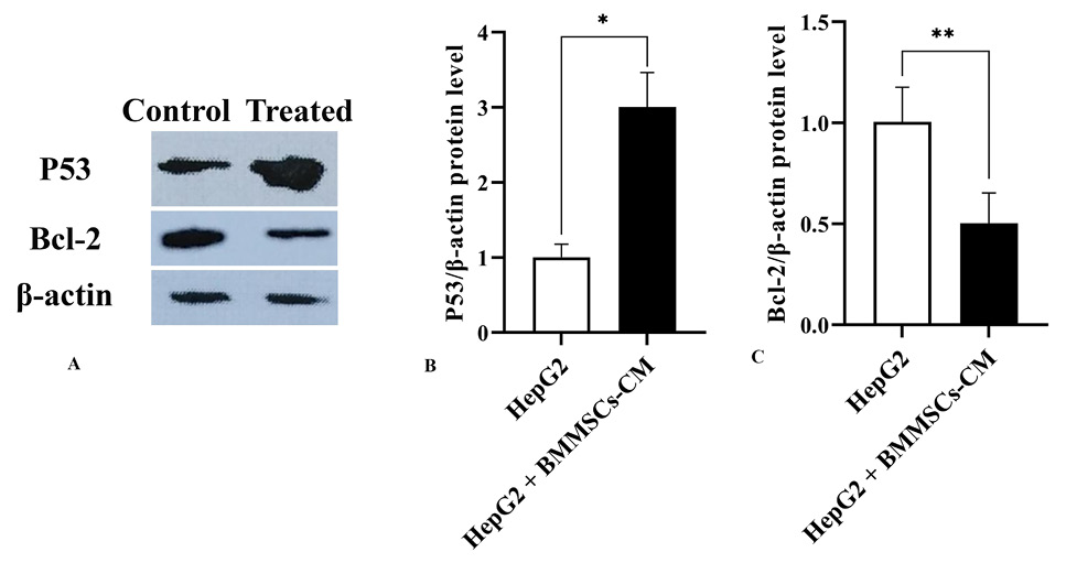
The effect of bone marrow mesenchymal stem cells-conditioned medium (BMMSCs-CM) on the hepatocellular carcinoma cell line HepG2 protein expression of P53 and Bcl-2 b western blotting. The data shows western bolt bands of untreated HepG2 cell group (control), and the treated HepG2 cell group with 100% BMMSC-CM for 72 h for P53 and Bcl-2 protein expression with β-actin as the house keeping protein (A), and the statistical analysis for P53 (B) Bcl-2 (C) proteins expression. Values are expressed as mean±SD, (n=3), the treated group (HepG2=BMMSC-CM) was compared to the control group. (*P<0.05, **P<0.01).
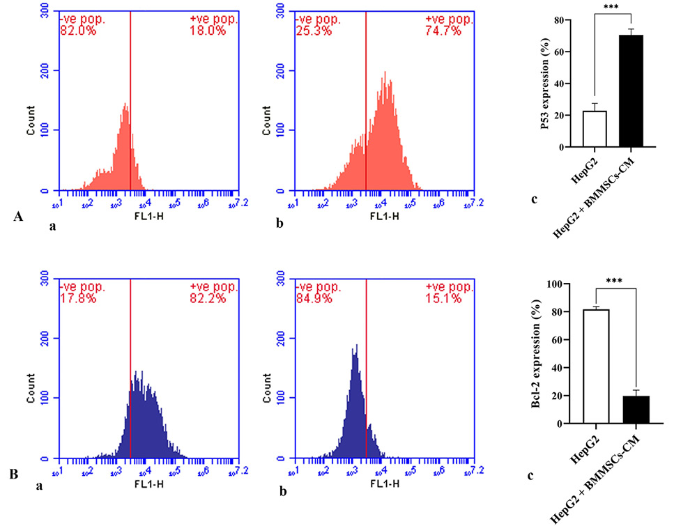
The effect of bone marrow mesenchymal stem cells-conditioned medium (BMMSCs-CM) on the hepatocellular carcinoma cell line HepG2 protein expression of P53 (A) and Bcl-2 (B) by floecytometry. The data shows the untreated HepG2 cell group (control), for P53 (Aa) and Bcl-2 (Ba) protein expression, and the statistical analysis for P53 (Ac) Bcl-2 (Bc) proteins expression. Values are expressed as mean±SD, (n=3), the treated group (HepG2=BMMSC-CM) was compared to the control group. (*P<0.05, **P<0.01).
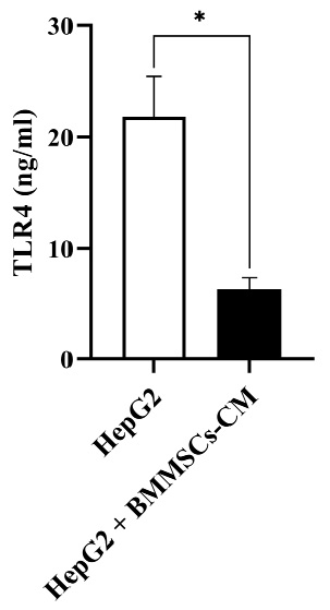
The influence of bone marrow mesenchymal stem cells-conditioned medium (BMMSCs-CM) on Toll-like receptor 4 (TLR4) concentration in the hepatocellular carcinoma cell line HepG2 by ELISA. The data shows the quantitative values of TLR4 concentration for untreated HepG2 cell group (control) and treated HepG2 cell groups with 100% BMMSC-CM for 72 h. Values are expressed as mean±SD, (n=3), the treated group (HepG2=BMMSC-CM) was compared to the control group (HepG2). (*P<0.05).


