
Description of Seven New Species and One New Record of Plant-Parasitic Nematodes (Nematoda: Tylenchida) Associated with Economically Important Crops of Kashmir Valley, Jammu and Kashmir (Part-1 of the series)
Zafar Ahmad Handoo1*, Mihail Radu Kantor1 and Ekramullah Khan2
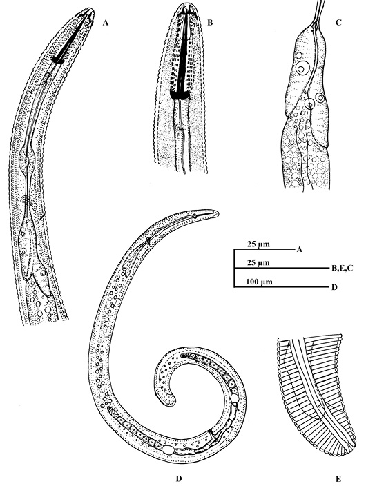
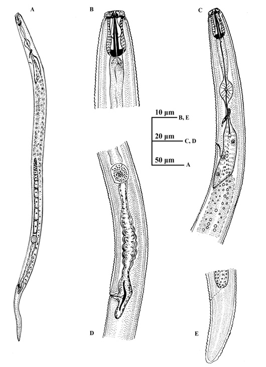
Helicotylenchus siddiqii sp. nov. A: Oesophageal region of female; B: Anterior end of female; C: Basal bulb of oesophagus; D: Entire female; E: Tail region female.

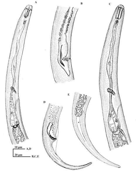
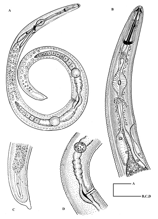
Helicotylenchus fotedariensis sp. nov. A: Entire female; B: Oesophageal region of female; C: Tail region of female; D: Vulval region showing gonad.
Helicotylenchus mushtaqi sp. nov. A: Anterior end of female; B: Entire female; C: Oesophageal region of female; D: Vulval region showing gonads; E: Tail region of female.
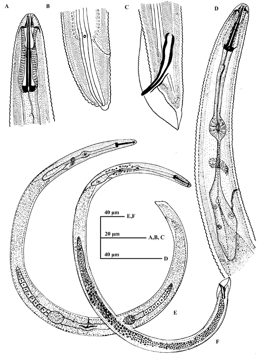
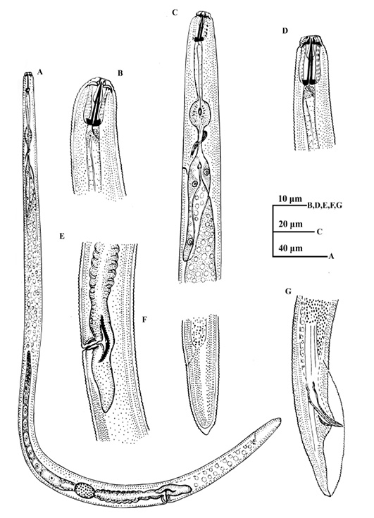
Pratylenchus ekrami Bajaj and Bhatti, 1984. A: Entire female; B: Anterior end of female; C: Oesophageal region of female; D: Anterior end of male; E: Vulval region showing anterior gonad; F: Tail region of female; G: Tail region of male.

Pratylenchus badawariensis sp. nov. A: Entire female; B: Anterior end of female; C: Oesophageal region of female; D: Vulval region of female showing anterior gonad; E: Tail region of female.
Boleodorus seshadri sp. nov. A: Oesophageal region of male; B: Vulval region showing posterior uterine sac; C: Anterior end of female; D: Tail region of male; E: Tail region of female showing phasmid.
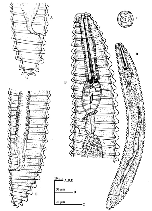
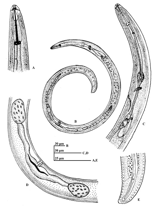
Macroposphora iqbali sp. nov. A: Female tail lateral view; B: Anterior end of female; C: En-face view of female; D: Entire female; E: Female tail lateral view.
