
Retrospective Seasonal Parasitological Survey on Prevalence and Epidemiological Determinants of Ectoparasitic Infestations in Dogs and Cats of Damietta, Egypt


Retrospective Seasonal Parasitological Survey on Prevalence and Epidemiological Determinants of Ectoparasitic Infestations in Dogs and Cats of Damietta, Egypt
Eman M. Aboelela1, Mohamed A. Sobieh2, Eman M. Abouelhassan3, Doaa S. Farid4, Essam S. Soliman2*

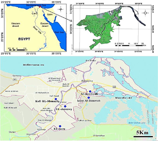
Geographical map for Damietta governorate, Egypt (In: Elgammal M, Ali RR, Samra RA. Assessing of heavy metal pollution in soils of Damietta governorate, Egypt. International Conference on Advances in Agricultural, Biological and Environmental Sciences (AABES-2014) Oct 15-16, 2014 Dubai).

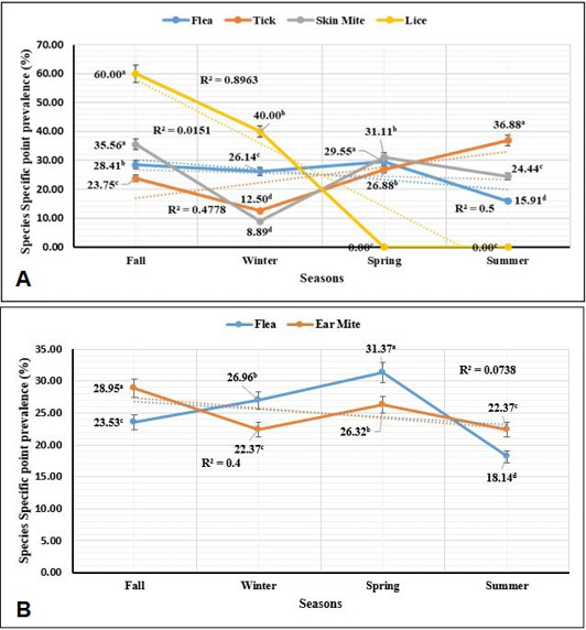
Species-specific point prevalence of different external parasites infesting dogs and cats concerning seasonal variation. A: External parasites infesting dogs. B: External parasites infesting cats.

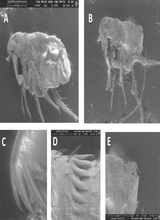
Scanning Electron microscopic picture of A: Ctenocephalides felis female, B: Ctenocephalides felis male, C: Spine 1 of genal ctenidium nearly equal to spine 2, D: Pronotal comb, and E: Posterior end male.
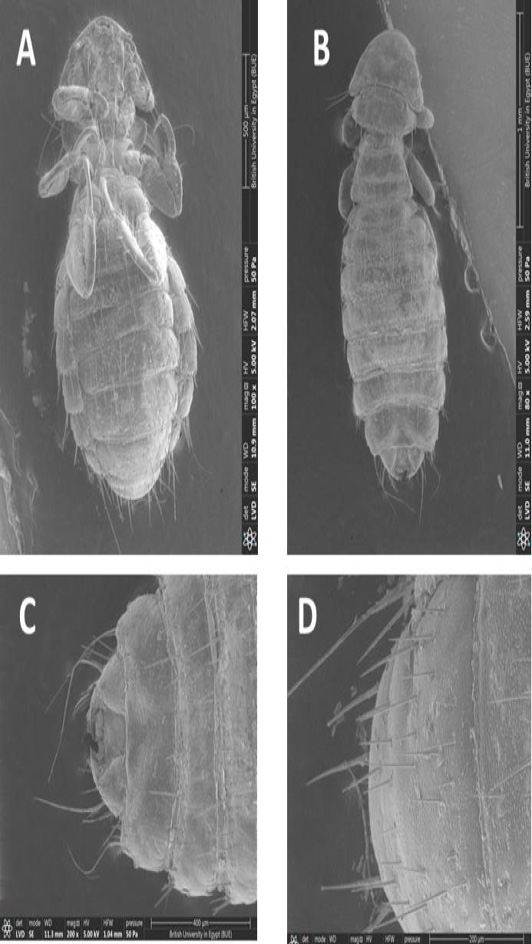
Scanning Electron microscopic picture of A: Heterodoxus spiniger male, B: Heterodoxus spiniger female, C: Posterior end female, and D: Posterior end male.

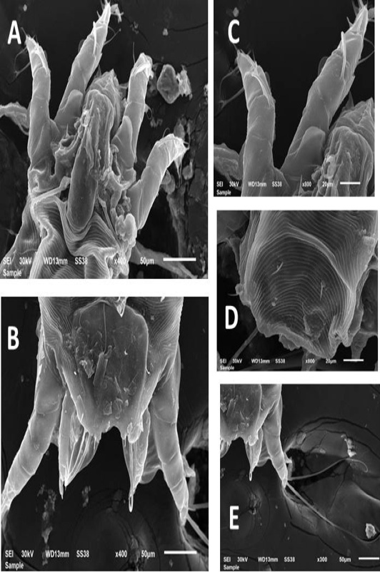
Scanning Electron microscopic picture of A: Otodectes cynotis, anterior end male, B: O. cynotis, posterior end male, C: anterior legs of O. cynotis male, D: finger-like projections present on the dorsum of O. cynotis, E: posterior legs of O. cynotis male.

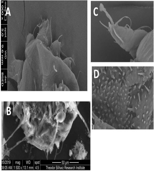
Scanning Electron microscopic picture of A: Sarcoptes scabiei, anterior end, B: S. scabiei, posterior end female, C: The un-segmented pedicels of anterior legs of S. scabiei female are terminate by a disk-like structure, D: Thorn like spines on the dorsum of Sarcoptic mite.
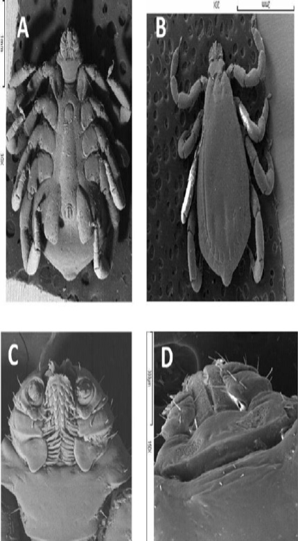
Scanning Electron microscopic picture of A: Rhipicephalus sanguineus ventral view, B: Rhipicephalus sanguineus dorsal view, C: Ventral view showing pedipalps and toothed hypostome, and D: Dorsal view showing hexagonal dorsal basis capituli.

